
Glycolysis - Glucose Breakdown
Introduction to Glycolysis
Glycolysis is the central pathway of glucose catabolism and one of the most important metabolic pathways in biology. Nearly every organism on Earth relies on some form of glycolysis to obtain energy from glucose. In this pathway, a single 6-carbon glucose molecule is converted into two 3-carbon pyruvate molecules through a series of 10 enzyme-catalyzed reactions. Glycolysis occurs in the cytosol and can operate under both aerobic and anaerobic conditions. Because the pathway does not directly require oxygen, it can continue producing ATP even when mitochondrial respiration is limited or unavailable.
Glycolysis can be divided into two major phases: the energy investment phase and the energy payoff phase. During the energy investment phase, the cell uses two ATP molecules to phosphorylate and activate glucose intermediates. These phosphorylation reactions also help trap metabolites within the cell, prepare the carbon skeleton for later cleavage, and prime the molecule for subsequent energy production. During the energy payoff phase, each glucose molecule gives rise to four ATP molecules and two NADH molecules through a series of oxidation and substrate-level phosphorylation reactions. Because two ATP molecules were consumed during the investment phase, glycolysis yields a net gain of two ATP per glucose molecule.
One reason glycolysis is often one of the first metabolic pathways taught in biochemistry is that it introduces many of the concepts that appear repeatedly throughout metabolism. As I progressed through my undergraduate biochemistry courses, I quickly realized that understanding glycolysis provides an essential foundation for understanding various other metabolic principles. Concepts such as energy coupling, pathway regulation, ATP production, redox chemistry, and metabolic flux all appear within this single pathway and reemerge throughout the rest of metabolism.
One of the major concepts throughout glycolysis is energy coupling. Endergonic reactions that would not proceed spontaneously are driven forward by coupling them to highly exergonic processes, most commonly ATP hydrolysis. Another concept theme is pathway regulation. Glycolysis must respond dynamically to cellular energy demands, nutrient availability, oxygen levels, and hormonal signals. As a result, several glycolytic enzymes serve as important regulatory control points that adjust pathway flux according to the metabolic state of the cell.
As we work through the pathway step by step, it is worth keeping the bigger picture in mind. Glycolysis is not simply a sequence of ten reactions to memorize. It is a coordinated process that extracts energy from glucose while producing metabolic intermediates that connect to many other pathways throughout the cell.
Energy Investment Phase of Glycolysis
STEP 1: HEXOKINASE
Reaction & Classifications
The first step of glycolysis is catalyzed by hexokinase, which phosphorylates glucose to form glucose-6-phosphate (G6P). In this reaction, ATP donates its terminal (γ) phosphate group to the hydroxyl group on carbon 6 of glucose, producing glucose-6-phosphate and ADP. The reaction is classified as a phosphorylation.
Hexokinase belongs to the transferase enzyme class, specifically the kinase subclass, because it transfers a phosphate group from ATP to a substrate. This reaction represents the first ATP investment of glycolysis and serves as the entry point for glucose into the pathway.
Overall Reaction: Glucose + ATP → Glucose-6-Phosphate + ADP
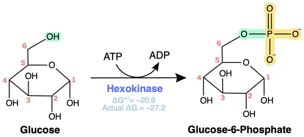
Figure 3. Hexokinase catalyzes the ATP-dependent phosphorylation of glucose to form glucose-6-phosphate, trapping glucose within the cell and initiating glycolysis.
Mechanistic Description
Hexokinase uses acid-base catalysis to perform a phosphoryl transfer through a direct nucleophilic substitution mechanism, coupling ATP hydrolysis to irreversible glucose phosphorylation. This reaction begins when the hydroxyl oxygen on C6 is deprotonated by an active site amino, allowing a nucleophilic lone pair of the C6 oxygen to attack the electrophilic γ-phosphate of ATP. As the new phosphate-oxygen bond forms, the structure passes through a pentavalent transition state before the bond between the γ-phosphate and ADP is broken, releasing ADP as the leaving group. In my undergrad biochem courses, my professor always referred to this state where glucose and ATP and complexed by a phosphate with 5 bonds the “pentavalent transition state.” Although there are probably more technical terms for this complex, out of habit and good memories I will be referring to this as the “pentavalent transition state” throughout my lessons.
Magnesium ions (Mg²⁺) are required for catalysis because they stabilize the negatively charged phosphate groups and properly orient ATP within the active site. By reducing electrostatic repulsion between the phosphate groups, Mg²⁺ promotes substrate binding and efficient phosphate transfer. This stabilization is especially important during formation of the pentavalent transition state, where it helps lower the activation energy of the reaction. The use of Mg²⁺ for catalysis is a common feature of most kinases involved in metabolism.

Figure 4. Hexokinase/glucokinase catalyzes the transfer of the γ-phosphate from ATP to glucose, producing glucose-6-phosphate through a pentavalent phosphoryl transition state.
Thermodynamics
The hexokinase reaction is strongly exergonic and essentially irreversible under physiological conditions.
ΔG°′ ≈ −20.9 kJ/mol | ΔG ≈ −27.2 kJ/mol
Due to this large negative free energy change, the reaction operates far from equilibrium within the cell and strongly favors formation of glucose-6-phosphate.
Regulation
Hexokinase is regulated primarily through feedback inhibition by its product, glucose-6-phosphate. When glucose-6-phosphate accumulates, it binds allosterically to hexokinase and decreases enzyme activity. This prevents excessive phosphorylation of glucose when downstream pathways are already saturated. In this way, product inhibition helps align the rate of glucose phosphorylation to the cell’s metabolic state, preventing unnecessary ATP expenditure and excessive accumulation of glucose-6-phosphate when downstream pathways are already operating at capacity. *Note that this regulation only applies to hexokinase, not glucokinase (a hexokinase isozyme) which is discussed below.
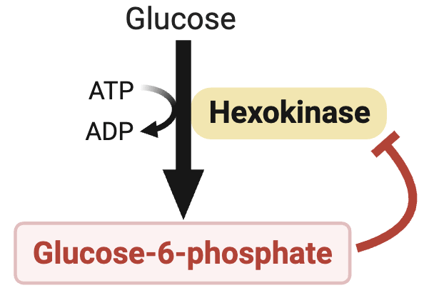
Figure 5. Hexokinase is inhibited by its product, glucose-6-phosphate, providing negative feedback that limits glucose phosphorylation when intracellular glucose-6-phosphate accumulates.
Other Notes
Induced-Fit Conformational Change
Hexokinase is a classic example of induced-fit enzyme catalysis. Prior to substrate binding, the enzyme exists in a relatively open conformation. When glucose binds, large conformational changes occur that cause the two domains of the enzyme to close around the substrate. This conformational change serves several purposes. It precisely positions glucose and ATP for phosphate transfer, stabilizes the transition state, and largely excludes water from the active site. Excluding water is particularly important because it prevents ATP from undergoing wasteful hydrolysis by water acting as a competing nucleophile. As a result, ATP hydrolysis becomes tightly coupled to glucose phosphorylation rather than occurring independently.
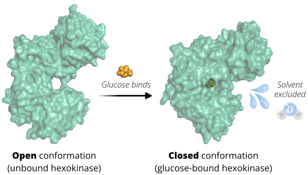
Figure 6. Glucose-induced conformational change in hexokinase. In the absence of substrate, hexokinase primarily exists in an open apoenzyme conformation with an exposed active-site cleft. Binding of glucose triggers a large induced-fit conformational change that closes the active site around the substrate, excludes solvent, and positions catalytic residues for efficient phosphoryl transfer from ATP.
PDB: 1IG8 (open, unbound) 1BDG (closed, bound)
Trapping Glucose Within the Cell
One of the most important consequences of this reaction is that it effectively traps glucose inside the cell. Glucose can enter and leave many cells through GLUT transporters, which are bidirectional. However, the addition of a negatively charged phosphate group dramatically changes the molecule’s properties. Glucose-6-phosphate cannot readily cross the plasma membrane and is not recognized by GLUT transporters. By rapidly converting incoming glucose into glucose-6-phosphate, intracellular free glucose concentrations remain low. This helps maintain a concentration gradient that favors continued glucose uptake from the bloodstream. In this way, hexokinase indirectly promotes glucose transport into cells while simultaneously committing glucose to intracellular metabolism.

Figure 7. Phosphorylation of glucose to glucose-6-phosphate traps glucose within the cell because glucose-6-phosphate is not transported by GLUT proteins, maintaining a concentration gradient that favors continued glucose uptake.
Hexokinase Isozymes and Glucokinase
Although several hexokinase isozymes exist, the most important comparison is between hexokinase and glucokinase. Most tissues express hexokinase, which has a low Km (high affinity) for glucose, meaning it can efficiently phosphorylate glucose even when blood glucose concentrations are low. Hexokinase also has a relatively low Vmax, so its maximum rate of glucose phosphorylation is limited, and it is inhibited by glucose-6-phosphate. These properties make hexokinase well suited for tissues that require a constant supply of glucose, such as the brain and skeletal muscle. Its high affinity ensures that cells can continue trapping and using glucose even during fasting or between meals, while product inhibition prevents glucose-6-phosphate from accumulating when the cell’s energy needs have already been met. This helps conserve ATP and allows glucose to remain available for other tissues that need it.
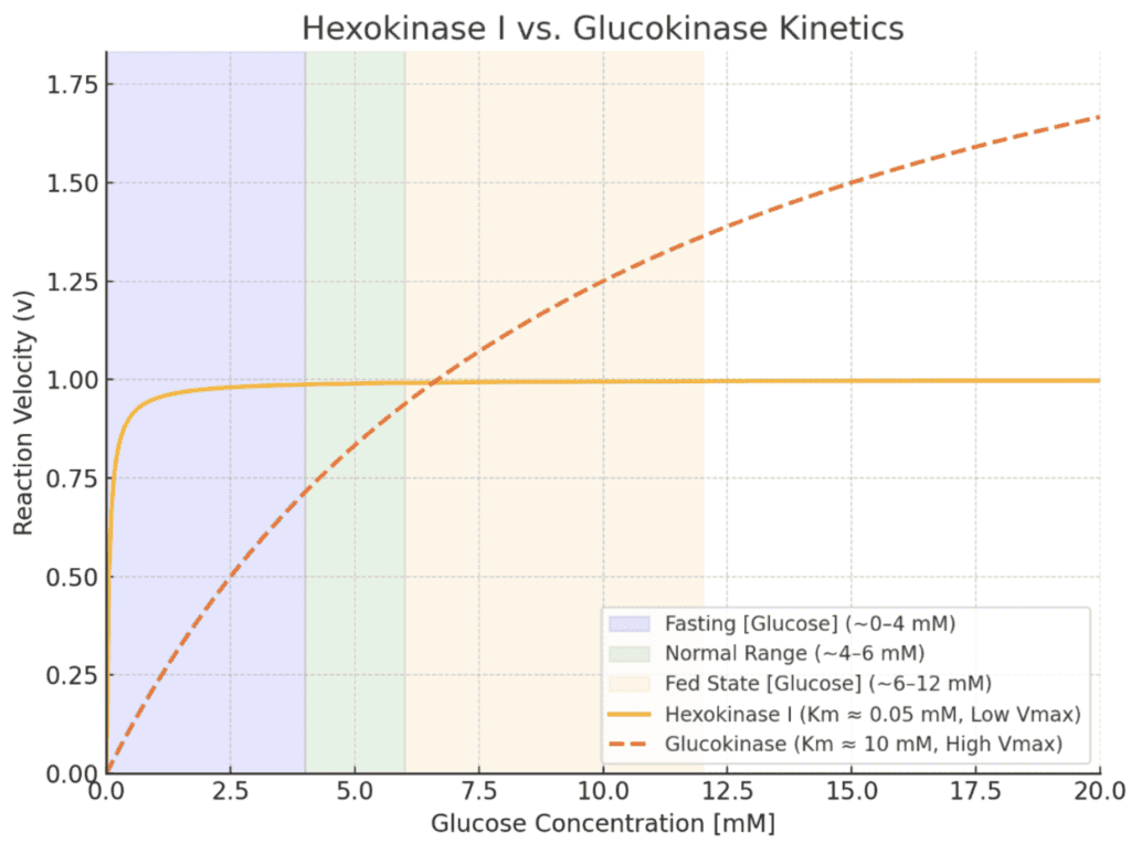
Figure 8. Text
In contrast, glucokinase is expressed primarily in the liver and pancreatic β-cells. Glucokinase has a high Km (low affinity) for glucose, meaning its activity increases substantially only when blood glucose concentrations rise after a meal. It also has a high Vmax, allowing it to phosphorylate large amounts of glucose when glucose is abundant, and it is not inhibited by glucose-6-phosphate. These properties make glucokinase well suited for the liver’s role in maintaining blood glucose levels. After a carbohydrate-rich meal, the liver can rapidly remove excess glucose from the bloodstream, convert it to glucose-6-phosphate, and either store it as glycogen or use it for metabolism. Because glucokinase is relatively inactive at low blood glucose concentrations, the liver does not compete with other tissues for glucose during fasting, allowing organs such as the brain and red blood cells to receive priority access to this essential fuel. In pancreatic β-cells, glucokinase acts as the cell’s glucose sensor. As blood glucose levels rise, increased glucokinase activity helps trigger insulin secretion, allowing the body to remove glucose from the bloodstream and restore normal blood glucose levels.
STEP 2: Phosphoglucose Isomerase
Reaction & Classifications
The second step of glycolysis is catalyzed by phosphoglucose isomerase (PGI), which converts glucose-6-phosphate (G6P) into fructose-6-phosphate (F6P). This reaction is classified as an isomerization, shifting the carbonyl group from carbon 1 to carbon 2, converting an aldohexose into a ketohexose.
Phosphoglucose isomerase belongs to the isomerase enzyme class because it catalyzes an intramolecular rearrangement without changing the overall molecular formula. More specifically, the reaction is classified as an aldose-ketose isomerization.
Overall Reaction: Glucose-6-Phosphate ⇌ Fructose-6-Phosphate
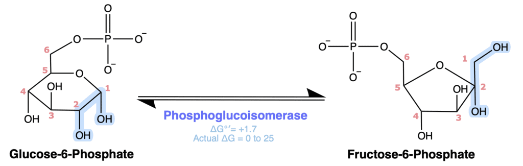
Figure 9. Phosphoglucose isomerase reversibly converts glucose-6-phosphate to fructose-6-phosphate by shifting the carbonyl group from C1 to C2.
Mechanistic Description
Phosphoglucose isomerase catalyzes a reversible aldose-ketose isomerization through a cis-enediol(ate) intermediate without changing the carbon skeleton of the sugar. The reaction begins with ring opening of glucose-6-phosphate to generate its linear aldehyde form. Once the sugar is in its open-chain conformation, active site residues catalyze proton transfer to form a cis-enediol(ate) intermediate. Specifically, a proton is abstracted from C2, and the resulting electrons form a C1-C2 double bond as the π electrons of the C1 carbonyl shift onto the oxygen, which is protonated to produce the cis-enediol(ate) intermediate. Formation of this intermediate allows the carbonyl group to shift from carbon 1 (aldose) to carbon 2 (ketose) without altering the carbon skeleton. Next, the hydroxyl group at C1 is deprotonated, allowing its lone pair to reform the carbonyl while the π electrons of the C1-C2 double bond shift onto C2, which is then protonated to form the hydroxyl group of fructose-6-phosphate. Finally, the C5 hydroxyl group acts as a nucleophile and attacks the electrophilic carbonyl carbon at C2, while the π electrons of the C=O bond shift onto the oxygen to form an alkoxide intermediate. Protonation of the alkoxide produces the cyclic hemiketal, forming the furanose ring of fructose-6-phosphate. The defining feature of this mechanism is the transient formation of the cis-enediol(ate) intermediate, which enables interconversion between the aldose and ketose forms of the sugar while preserving the carbon skeleton.

Figure 10. Phosphoglucose isomerase converts glucose-6-phosphate to fructose-6-phosphate by reversible ring opening, formation of a cis-enediol intermediate, and ring closure.
Thermodynamics
The phosphoglucose isomerase reaction is near equilibrium under physiological conditions. ΔG°′ ≈ +1.7 kJ/mol. Although the standard free energy change is slightly positive, the actual cellular ΔG is typically close to zero and often slightly negative because fructose-6-phosphate is continuously consumed by phosphofructokinase-1 in the next step of glycolysis. As a result, reaction equilibrium shifts to favor product formation and forward flux through the pathway.
Regulation
Phosphoglucose isomerase is not considered a major regulatory enzyme of glycolysis. Because the reaction operates close to equilibrium, its direction is determined primarily by substrate and product concentrations rather than by extensive allosteric regulation. Consequently, pathway flux through this step is largely controlled by the enzymes that precede and follow it, particularly hexokinase and phosphofructokinase-1.
Other Notes
Why Fructose Is Needed
I’ve often heard students ask why forming the fructose ring is necesarry, why not just keep the glucose pyranose form? The answer becomes clear when examining the later aldolase reaction (step 4). The 6-carbon glucose molecule must eventually be split into two 3-carbon molecules. Converting glucose-6-phosphate into fructose-6-phosphate moves the carbonyl group to the center of the molecule, creating a structure that can be cleaved into two phosphorylated 3-carbon intermediates of comparable energy content. This seemingly small rearrangement is therefore essential for the overall design of the pathway.
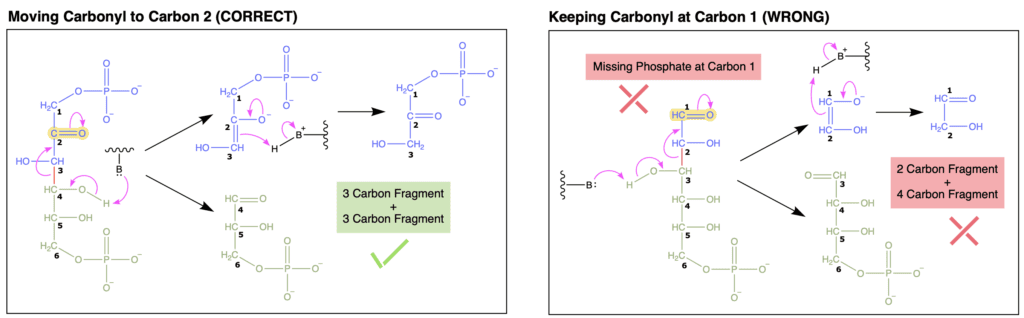
Figure 11. text.
Reversibility and Metabolic Flexibility
Since this reaction is readily reversible, phosphoglucose isomerase participates in several pathways beyond glycolysis. During gluconeogenesis, for example, the enzyme catalyzes the reverse conversion of fructose-6-phosphate back to glucose-6-phosphate. This reversibility is a common feature of many near-equilibrium reactions in metabolism. These reactions can proceed in either direction depending on the needs of the cell.
STEP 3: Phosphofructokinase-1 (PFK-1)
Reaction & Classifications
The third step of glycolysis is catalyzed by phosphofructokinase-1 (PFK-1), one of the most important regulatory enzymes in metabolism. In this reaction, ATP donates a phosphate group to the hydroxyl group on carbon 1 of fructose-6-phosphate, producing fructose-1,6-bisphosphate (F1,6BP).
Like hexokinase, PFK-1 belongs to the kinase subclass of the transferase enzyme family because it transfers a phosphate group from ATP to a substrate. The reaction is classified as a phosphorylation.
This reaction is the second ATP-consuming step of glycolysis and marks the first committed step of the pathway. Once this reaction occurs, the glucose molecule is committed to continuing through glycolysis.
Overall Reaction: Fructose-6-Phosphate + ATP → Fructose-1,6-Bisphosphate + ADP
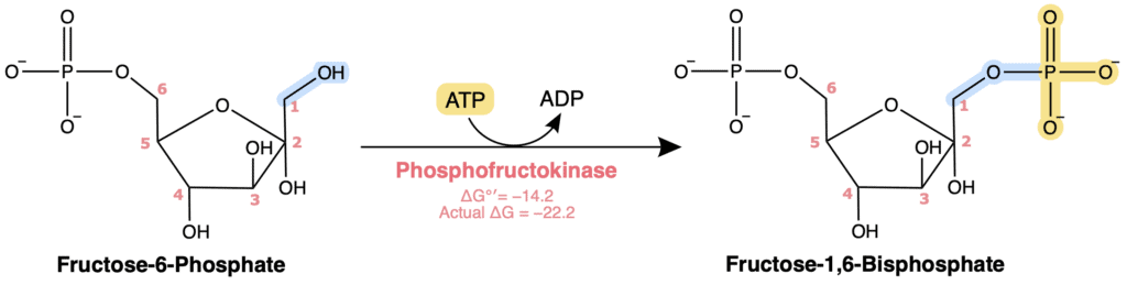
Figure 12. Phosphofructokinase-1 catalyzes the ATP-dependent phosphorylation of fructose-6-phosphate to produce fructose-1,6-bisphosphate, the committed step of glycolysis.
Mechanistic Description
Phosphofructokinase-1 catalyzes irreversible phosphoryl transfer from ATP to fructose-6-phosphate, committing the sugar to glycolysis. This reaction begins when an active site residue acts as a base to abstract the proton from the C1 hydroxyl group, allowing the resulting oxygen lone pair to attack the electrophilic γ-phosphorus atom of ATP. Formation of this new oxygen-phosphorus bond transiently generates a pentavalent transition state as the electrons of the P=O bond shift onto the oxygen. The electrons then collapse back to reform the P=O bond, while the electrons of the phosphoanhydride bond shift onto the bridging oxygen, releasing ADP as the leaving group. Finally, the active site residue protonates the bridging oxygen, completing phosphoryl transfer and producing fructose-1,6-bisphosphate. The defining feature of this mechanism is direct phosphoryl transfer through a pentavalent transition state. This irreversible phosphorylation commits the substrate to glycolysis and represents the pathway’s primary rate-limiting step.
Like most kinases, magnesium ions (Mg²⁺) are required for catalysis to help stabilize the negative charges on the phosphates and orient ATP within the active site. Also similar to hexokinase, PFK-1 uses solely acid-base catalysis and does not form a covalent enzyme-substrate intermediate.

Figure 13. Phosphofructokinase-1 transfers the γ-phosphate from ATP to fructose-6-phosphate, producing fructose-1,6-bisphosphate through a pentavalent phosphoryl transition state.
Thermodynamics
The PFK-1 reaction is strongly exergonic and essentially irreversible under physiological conditions.
ΔG°′ ≈ −14.2 kJ/mol | ΔG ≈ −22.2 kJ/mol
Although the standard free energy change is somewhat less negative than that of the hexokinase reaction, the actual cellular ΔG is typically much more negative because fructose-6-phosphate concentrations are maintained well below equilibrium levels. As a result, the reaction operates far from equilibrium and proceeds strongly in the forward direction.
This step holds a unique position within glycolysis because it is considered the committed step of the pathway. Glucose-6-phosphate and fructose-6-phosphate can still be diverted into alternative pathways such as glycogen synthesis or the pentose phosphate pathway. Once fructose-1,6-bisphosphate is formed, however, the carbon skeleton is effectively committed to glycolytic metabolism.
Because it controls entry into the remainder of the pathway, PFK-1 serves as the major rate-limiting and regulatory step of glycolysis. Consequently, many signals that reflect the metabolic state of the cell converge on this enzyme.
Regulation
PFK-1 is extensively regulated through allosteric interactions that allow glycolytic flux to respond rapidly to changes in cellular energy status.
ATP serves both as a substrate and as an allosteric inhibitor. At low concentrations, ATP participates normally in catalysis. However, when ATP concentrations become high, ATP binds to a separate regulatory site and decreases enzyme activity. This regulation allows the cell to slow glycolysis when energy supplies are already abundant.
AMP is a potent allosteric activator of PFK-1. Rising AMP levels indicate that ATP has been consumed and that cellular energy stores are declining. Activation of PFK-1 under these conditions increases glycolytic flux and promotes ATP production. ADP can also contribute to activation, although AMP generally serves as the more sensitive indicator of cellular energy status.
Citrate acts as an allosteric inhibitor of PFK-1. Elevated citrate levels signal that the TCA cycle is already adequately supplied with carbon and reducing equivalents. Under these conditions, additional glycolytic input is unnecessary, and PFK-1 activity decreases. Citrate therefore provides an important link between glycolysis and mitochondrial metabolism.
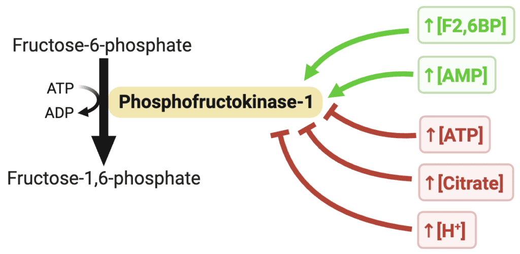
Figure 14. Regulation overview for phosphofructokinase-1 (PFK-1). Fructose-2,6-bisphosphate and AMP activate the enzyme, whereas ATP, citrate, and elevated H⁺ inhibit its activity, making PFK-1 the primary regulatory enzyme of glycolysis.

Figure 15. text
One of the most powerful activators of PFK-1, particularly in the liver, is fructose-2,6-bisphosphate (F2,6BP). F2,6BP increases the affinity of PFK-1 for fructose-6-phosphate while simultaneously reducing the inhibitory effects of ATP. As a result, glycolysis can remain active even when ATP concentrations are relatively high. The concentration of fructose-2,6-bisphosphate is controlled by the bifunctional enzyme PFK-2/FBPase-2, which plays a central role in coordinating glycolysis and gluconeogenesis. We will revisit this regulatory system in greater detail when discussing hormonal control of metabolism.
Other Notes
The Most Important Regulatory Enzyme of Glycolysis
If there is one glycolytic enzyme worth remembering, it is PFK-1. While hexokinase initiates glycolysis and pyruvate kinase completes it, PFK-1 serves as the primary decision point of the pathway. More than any other glycolytic enzyme, PFK-1 determines whether glucose will continue through glycolysis or be directed elsewhere in metabolism. For this reason, many biochemistry courses devote substantial attention to understanding PFK-1 regulation.
Structural Basis of Allosteric Regulation
PFK-1 functions as a homotetramer composed of four identical subunits. The enzyme can adopt two major conformational states: a less active T (tense) state and a more active R (relaxed) state. Allosteric activators such as AMP and fructose-2,6-bisphosphate stabilize the R state and promote catalysis. In contrast, inhibitors such as ATP and citrate stabilize the T state and reduce catalytic activity. These conformational transitions allow PFK-1 to simultaneously integrate multiple metabolic signals and adjust the rate of glycolytic flux accordingly.
Figure 16. text
STEP 4: Aldolase
Reaction & Classifications
The fourth step of glycolysis is catalyzed by aldolase, which cleaves fructose-1,6-bisphosphate into two 3-carbon intermediates: dihydroxyacetone phosphate (DHAP) and glyceraldehyde-3-phosphate (GAP). This reaction represents the first carbon-carbon bond cleavage in glycolysis and marks the point at which the 6-carbon glucose skeleton is divided into two smaller molecules.
Aldolase belongs to the lyase enzyme class because it cleaves a carbon-carbon bond without hydrolysis or oxidation. More specifically, the reaction is classified as an aldol cleavage.
Although the products differ structurally, each contains three carbons and one phosphate group. These molecules will ultimately be converted into glyceraldehyde-3-phosphate before entering the energy payoff phase of glycolysis.
Overall Reaction: Fructose-1,6-Bisphosphate ⇌ Dihydroxyacetone Phosphate + Glyceraldehyde-3-Phosphate
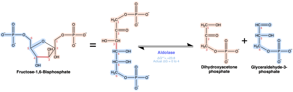
Figure 17. Aldolase reversibly cleaves fructose-1,6-bisphosphate into the two three-carbon intermediates dihydroxyacetone phosphate and glyceraldehyde-3-phosphate.
Mechanistic Description
Aldolase catalyzes cleavage of the bond between carbons 3 and 4 of fructose-1,6-bisphosphate. In animals and plants, this reaction is carried out by class I aldolases, which utilize covalent catalysis.
The reaction begins with ring opening of fructose-1,6-bisphosphate to generate its linear ketone form. The ε-amino group of an active site lysine residue then acts as a nucleophile and attacks the electrophilic carbonyl carbon at C2, while the π electrons of the C=O bond shift onto the oxygen. Subsequent proton transfer and elimination of water produce a covalent Schiff base (imine) intermediate, which acts as an electron sink that stabilizes negative charge development during subsequent bond cleavage. For simplicity, the arrows shown in pink with an asterisk (*) in Figure X depicts a simplified version of the imine formation mechanism. The complete proton transfer steps are described in the side note below. An active site residue then abstracts the proton from the C4 hydroxyl group, allowing the resulting oxygen lone pair to reform the carbonyl while the electrons of the C3-C4 bond shift to cleave the C3-C4 bond. This retro-aldol cleavage releases glyceraldehyde-3-phosphate and leaves the remaining three-carbon fragment covalently attached to the lysine residue as a resonance-stabilized carbanion. Next, electron resonance rearrangement of the carbanion reforms the Schiff base and places the lone pair on C3, which is then protonated. Finally, hydrolysis of the imine releases dihydroxyacetone phosphate and regenerates the free lysine residue, preparing the enzyme for another catalytic cycle. The defining feature of this mechanism is formation of the Schiff base intermediate, which acts as an electron sink to stabilize the high-energy carbanion intermediate and enable retro-aldol cleavage.
SIDE NOTE:
One thing I found especially confusing when learning this mechanism was how many different versions of it appear in textbooks and online. From what I’ve seen, most sources use a simplified aldolase mechanism that omits the individual steps involved in forming the imine (Schiff base). This is common because proton transfer and dehydration steps are often left out of organic mechanisms unless they are directly relevant to the concept being taught. Readers are generally expected to understand that these steps occur, even if they are not shown. Including every intermediate can also make the mechanism much longer without adding much to the overall concept.
This simplified version is the one I use in Figure X, where the condensed steps are marked with an asterisk (*). However, since I’m an organic chemistry enthusiast, and I know some of you are too, I’ve also included the complete mechanism for imine formation in Figure X for anyone interested in seeing every step.
Just remember that there are several acceptable ways to draw and simplify this mechanism. Be sure to check with your professor to see which version they expect you to know.
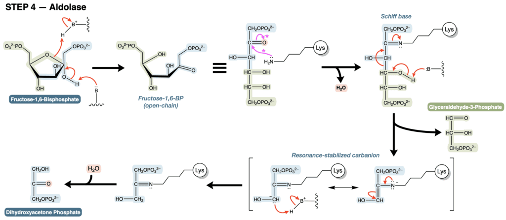
Figure 18. Aldolase catalyzes the cleavage of fructose-1,6-bisphosphate into glyceraldehyde-3-phosphate and dihydroxyacetone phosphate through formation of a Schiff base and a resonance-stabilized carbanion intermediate.
Thermodynamics
The aldolase reaction has a strongly positive standard free energy change.
ΔG°′ ≈ +23.8 kJ/mol
At first glance, this suggests that the reaction is thermodynamically unfavorable in the forward direction. However, the actual cellular ΔG is much closer to equilibrium because when glycolysis is active, there is a constant accumulation of upstream substrates and removal of products by downstream reactions to push the reaction in the forward direction.
Regulation
Aldolase is not considered a major regulatory enzyme of glycolysis. Like step 2, this reaction operates near equilibrium under physiological conditions, so its direction and rate are determined primarily by the concentrations of its substrates and products. As a result, flux through this step is largely controlled by regulatory enzymes elsewhere in the pathway.
Other Notes
Why Glucose Must Be Split
Up to this point, the preceding glycolytic reactions have served as preparatory steps, involving phosphorylation and isomerization, that convert glucose into a 6-carbon molecule capable of being cleaved into two phosphorylated 3-carbon molecules. This cleavage marks an important transition because it allows both halves of the original glucose molecule to be processed simultaneously through the same series of reactions. From this point forward, every remaining step occurs twice for each molecule of glucose, doubling the production of ATP and NADH during the payoff phase. 3-carbon molecules are also the idal size for the downstream reactions that ultimately produce pyruvate while capturing energy in forms the cell can use. This organization also improves metabolic efficiency because the cell only needs one set of enzymes to process both 3-carbon molecules rather than evolving separate pathways for each half of the original glucose molecule.
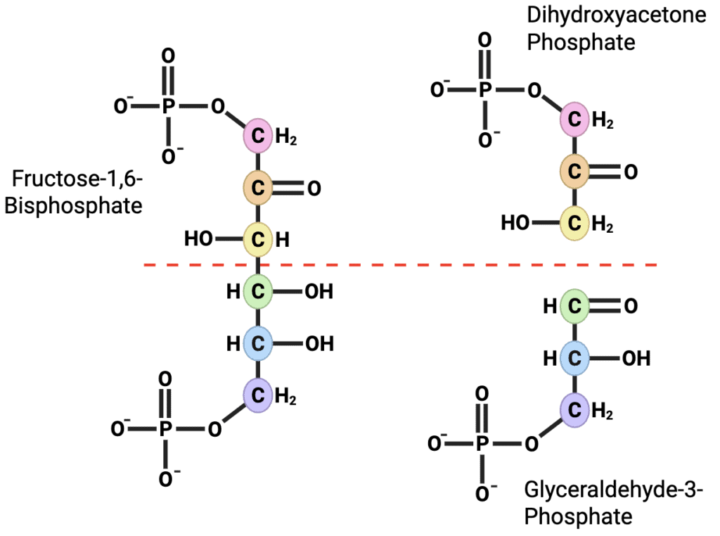
Figure 19. Aldolase cleaves fructose-1,6-bisphosphate between C3 and C4 to produce the two three-carbon intermediates dihydroxyacetone phosphate (C1-C3) and glyceraldehyde-3-phosphate (C4-C6).
Carbon-Tracking
Carbon tracking is an important part of understanding glycolysis, and it is especially important to pay attention to during the aldolase reaction. Because this is where the six carbon glucose molecule is split into two three carbon molecules, it can be easy to lose track of where each carbon ends up. At the end of the following TPI section, I’ll explain in detail how to track each carbon all the way to pyruvate.
Class I vs. Class II Aldolases
Two major classes of aldolase exist in nature. Class I aldolases, found in animals and plants, utilize Schiff base chemistry involving an active-site lysine residue. Class II aldolases, which are common in many microorganisms, do not form a Schiff base. Instead, they use divalent metal ions such as Zn²⁺ or Fe²⁺ to stabilize negatively charged reaction intermediates during bond cleavage. Although the mechanisms differ, both enzyme classes accomplish the same overall transformation.
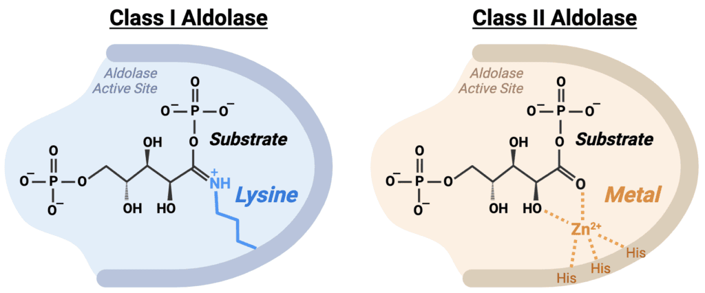
Figure 20. Comparison of the active sites of Class I and Class II aldolases. Class I aldolases form a covalent Schiff base intermediate with an active-site lysine residue, whereas Class II aldolases use a divalent metal ion, typically Zn²⁺ or Fe²⁺, to stabilize the reaction intermediate.
STEP 5: Triose Phosphate Isomerase (TPI)
Reaction & Classifications
The fifth step of glycolysis is catalyzed by triose phosphate isomerase (TPI), which interconverts dihydroxyacetone phosphate (DHAP) and glyceraldehyde-3-phosphate (GAP).
Triose phosphate isomerase belongs to the isomerase enzyme class because it catalyzes an intramolecular rearrangement without altering the overall molecular formula. More specifically, the reaction is classified as an aldose-ketose isomerization.
Although aldolase produces both DHAP and GAP, only glyceraldehyde-3-phosphate can proceed directly through the remainder of glycolysis. TPI therefore ensures that both three-carbon fragments generated from glucose can ultimately be metabolized.
Overall Reaction: Dihydroxyacetone Phosphate ⇌ Glyceraldehyde-3-Phosphate
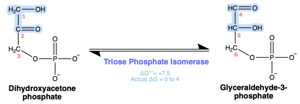
Figure 21. Triose phosphate isomerase reversibly interconverts dihydroxyacetone phosphate and glyceraldehyde-3-phosphate, allowing both three-carbon products to continue through glycolysis.
Mechanistic Description
Triose phosphate isomerase catalyzes a reversible aldose-ketose isomerization through a cis-enediol intermediate, rapidly interconverting the two triose phosphates. The reaction begins when an active site residue acts as a base to abstract the proton from C1 of DHAP. The resulting electrons form a C1=C2 double bond as the π electrons of the C2 carbonyl shift onto the oxygen, which is protonated to form a C2 hydroxyl group. After formation of this cis-enediol intermediate, the hydroxyl group at C1 is deprotonated, allowing its lone pair to reform the carbonyl while the π electrons of the C1=C2 double bond shift onto C2. Then, C2 is protonated to produce the aldehyde group of GAP, completing the reversible aldose-ketose isomerization. The defining feature of this mechanism is the formation of the cis-enediol intermediate, which enables rapid interconversion between dihydroxyacetone phosphate and glyceraldehyde-3-phosphate without altering the carbon skeleton.
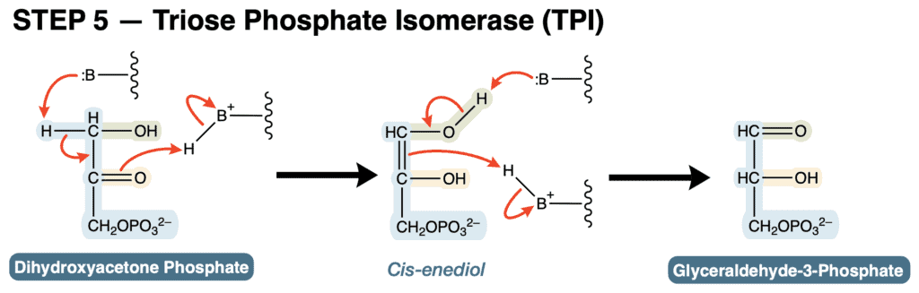
Figure 22. Triose phosphate isomerase reversibly interconverts dihydroxyacetone phosphate and glyceraldehyde-3-phosphate through a cis-enediol intermediate.
Thermodynamics
The triose phosphate isomerase reaction operates very close to equilibrium under physiological conditions.
ΔG°′ ≈ +7.5 kJ/mol
Despite the positive standard free energy change, the actual cellular ΔG is typically near zero because glyceraldehyde-3-phosphate is continuously consumed by glyceraldehyde-3-phosphate dehydrogenase (GAPDH) in the next step of glycolysis. This continual removal of GAP effectively pulls the reaction forward.
The physiological importance of this step cannot be overstated. Aldolase produces one molecule of DHAP and one molecule of GAP from each fructose-1,6-bisphosphate. If glycolysis could metabolize only GAP, half of the carbon atoms from glucose would be unable to continue through the pathway. TPI solves this problem by rapidly converting DHAP into GAP, allowing both three-carbon fragments to contribute to ATP and NADH production.As a result, all six carbons originally present in glucose can be fully utilized during the remainder of glycolysis.
Regulation
Triose phosphate isomerase is not considered a major regulatory enzyme of glycolysis. Again, this reaction operates near equilibrium so its direction depends primarily on substrate and product concentrations.
Other Notes
Near Catalytically Perfect Enzyme
TPI is frequently cited as one of the best examples of a catalytically perfect enzyme (very high Vmax). Its catalytic rate is limited primarily by substrate diffusion into the active site rather than by the chemical reaction itself. In practical terms, nearly every substrate molecule that successfully enters the active site is rapidly converted into product. For this reason, TPI is often used in biochemistry courses as a classic example of the upper limits of enzyme efficiency.
Figure X. text
Why the Reaction Matters
At first glance, converting one three-carbon molecule into another may seem relatively unimportant compared with the ATP-producing reactions that follow. However, this reaction effectively doubles the energy yield of glycolysis. Without TPI, only the GAP molecule produced by aldolase could continue through the pathway. The DHAP molecule would remain metabolically stranded, and only half of the original glucose molecule could be used for ATP production. By converting DHAP into GAP, TPI ensures that both halves of glucose contribute equally to the energy payoff phase.
The End of the Energy Inverstment Phase
The triose phosphate isomerase reaction marks the end of the energy investment phase of glycolysis. At this point, two ATP molecules have been consumed, and the original six-carbon glucose molecule has been converted into two molecules of GAP. From this stage onward, every remaining reaction in glycolysis occurs twice per original glucose molecule. This is an important point to remember when tracking ATP and NADH production. Beginning with the next reaction, the pathway effectively runs in parallel for two molecules of GAP, meaning that all subsequent yields must be multiplied by two when calculating the overall output of glycolysis.
An Important Carbon-Tracking Point
Many students find glycolysis much easier to understand once they begin tracking the carbon atoms through the pathway, especially as the investment phase comes to an end. As you work through these steps, I recommend either color coding each carbon or numbering them 1 through 6 so you can follow where every carbon from the original glucose molecule ends up.
One trick I find helpful is using the phosphate groups to orient the molecules. Before cleavage, fructose 1,6-bisphosphate has phosphates attached to carbons 1 and 6. After aldolase cleaves the molecule, glucose carbons 1, 2, and 3 become DHAP, while glucose carbons 4, 5, and 6 become GAP.
When drawing these molecules, I like to place the phosphate at the top of DHAP and at the bottom of GAP. This keeps the carbon numbers increasing from top to bottom in both molecules, making them much easier to follow. A simple way I remember this is that DHAP wears its phosphate like a hat, while GAP keeps its phosphate at the bottom because it has to “jump the gap.” This orientation is shown in Figure X.
Carbon tracking becomes a little trickier after the next step. Triose phosphate isomerase converts DHAP into GAP, so both products are now the same molecule. To match the orientation of the GAP produced directly by aldolase, I redraw the newly formed GAP with its phosphate at the bottom. As a result, the carbon numbering for the GAP derived from glucose carbons 1, 2, and 3 now appears to run in the opposite direction. In other words, if you are tracing the original glucose carbons, you will now number up one GAP molecule and down the other (Figure X).
From this point forward, both GAP molecules pass through exactly the same reactions. Although they are chemically identical, they still originate from different halves of the original glucose molecule. One GAP contains glucose carbons 1, 2, and 3, while the other contains glucose carbons 4, 5, and 6. Importantly, during the payoff phase the three carbon backbone of each molecule remains intact. No carbons are added, removed, or exchanged between the two molecules. The reactions only modify the functional groups attached to those carbons. This means that if you keep track of the carbon numbering at this point, you can follow every original glucose carbon all the way to the two pyruvate molecules produced at the end of glycolysis.
Personally, once both molecules have become GAP, I like to renumber each one from 1 to 3 for the remainder of glycolysis because it makes the payoff phase much easier to follow. Just remember that these are simply new reference numbers for convenience. The original glucose carbon numbers are still important because they tell you exactly where each carbon in the final pyruvate molecules came from.
Figure X. text.
STEP 6: Glyceraldehyde-3-Phosphate Dehydrogenase (GAPDH)
Reaction & Classifications
The sixth step of glycolysis is catalyzed by glyceraldehyde-3-phosphate dehydrogenase (GAPDH). In this reaction, glyceraldehyde-3-phosphate (GAP) is oxidized and phosphorylated to form 1,3-bisphosphoglycerate (1,3-BPG). This step marks the beginning of the energy payoff phase and is the first reaction in glycolysis that captures energy from glucose oxidation.
GAPDH belongs to the oxidoreductase enzyme class because it catalyzes an oxidation-reduction reaction. More specifically, the aldehyde group of GAP is oxidized while NAD⁺ is reduced to NADH. The reaction is also unusual because it incorporates inorganic phosphate (Pi) directly into the product rather than using ATP as the phosphate donor.
This reaction produces the first NADH molecules generated during glycolysis. Because two molecules of glyceraldehyde-3-phosphate are processed per original glucose molecule, the reaction occurs twice and ultimately produces two NADH per glucose.
Overall Reaction: Glyceraldehyde-3-Phosphate + NAD⁺ + Pi ⇌ 1,3-Bisphosphoglycerate + NADH + H⁺
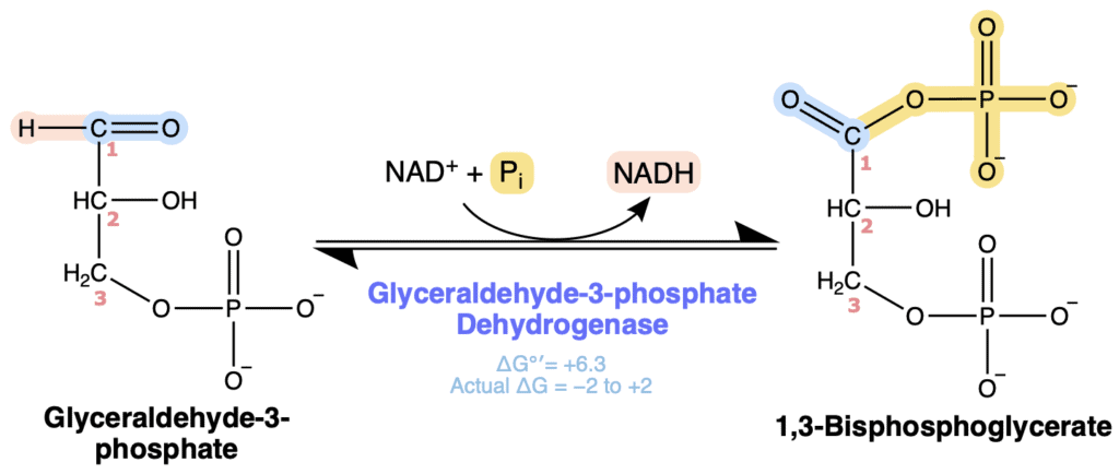
Figure X. Glyceraldehyde-3-phosphate dehydrogenase oxidizes glyceraldehyde-3-phosphate and incorporates inorganic phosphate to form 1,3-bisphosphoglycerate while reducing NAD⁺ to NADH.
Mechanistic Description
GAPDH utilizes both covalent catalysis and oxidation-reduction chemistry to conserve energy released during substrate oxidation. The reaction begins when an active site cysteine residue acts as a nucleophile and attacks the electrophilic aldehyde carbon of glyceraldehyde-3-phosphate, while the π electrons of the C=O bond shift onto the oxygen to form a covalent hemithioacetal intermediate. The intermediate is then oxidized as the carbonyl C=O bond reforms, transferring a hydride ion (:H⁻) from C1 to NAD⁺ to produce NADH. This oxidation step generates a high-energy thioester intermediate between the enzyme and substrate. Rather than allowing this energy to dissipate as heat, GAPDH captures and stores it within the thioester bond. Next, inorganic phosphate acts as a nucleophile and attacks the thioester carbonyl carbon, once again causing the π electrons of the C=O bond to shift onto the oxygen. The electrons then collapse back to reform the carbonyl, causing the carbon-sulfur bond to break and releasing the cysteine which is reprotonated, regenerating the enzyme and releasing 1,3-bisphosphoglycerate. The most important feature of the mechanism is the formation of the high-energy thioester intermediate. This allows the energy released during oxidation to be captured and stored within the acyl phosphate bond of 1,3-bisphosphoglycerate.

Figure X. Glyceraldehyde-3-phosphate dehydrogenase oxidizes glyceraldehyde-3-phosphate and couples NAD⁺ reduction with inorganic phosphate addition to form 1,3-bisphosphoglycerate.
Thermodynamics
The GAPDH reaction has a slightly positive standard free energy change.
ΔG°′ ≈ +6.3 kJ/mol | ΔG ≈ −2 to +2
Despite this positive ΔG°′, the actual cellular ΔG is typically close to zero or slightly negative because 1,3-BPG is rapidly consumed by phosphoglycerate kinase in the next step of glycolysis. As a result, the reaction proceeds efficiently in vivo.
Regulation
GAPDH is not considered a major allosteric regulatory enzyme of glycolysis. Instead, its activity depends largely on substrate availability and the cellular redox state, particularly the ratio of NAD⁺ to NADH. Because NAD⁺ serves as the electron acceptor for the reaction, glycolysis requires a continuous supply of oxidized NAD⁺ in order to proceed. Under aerobic conditions, NADH generated by glycolysis can be reoxidized through the electron transport chain. Under anaerobic conditions, alternative pathways such as lactate fermentation regenerate NAD⁺ and allow glycolysis to continue. If NAD⁺ becomes depleted, the GAPDH reaction slows dramatically, causing glycolytic flux to decrease regardless of the availability of glucose.
Other Notes
The First Energy-Conserving Reaction
Many students focus on the fact that GAPDH produces NADH, but the more important concept is that this reaction captures energy released during oxidation. If the aldehyde of GAP were oxidized directly to a carboxylic acid, much of the available energy would simply be lost as heat. Instead, GAPDH conserves this energy by forming the high-energy acyl phosphate bond of 1,3-BPG. This stored energy is immediately utilized in the next reaction to generate ATP.
The Importance of Thioesters
The thioester intermediate formed during catalysis introduces a strategy you’ll encounter many times throughout metabolism. As discussed earlier in the section on high-energy compounds, thioesters are considered energy rich because sulfur provides much less resonance stabilization than oxygen. As a result, thioesters are relatively unstable and have a large, negative free energy of hydrolysis. During the GAPDH reaction, the oxidation of GAP releases a substantial amount of free energy. Rather than allowing this energy to dissipate as heat, the enzyme temporarily conserves it by forming a high-energy thioester intermediate with an active site cysteine. Inorganic phosphate then replaces the cysteine, transferring the conserved energy into the high-energy acyl phosphate bond of 1,3-BPG, which is used to generate ATP in the next step of glycolysis. The same strategy of temporarily conserving energy in a thioester bond appears later with acetyl-CoA in the citric acid cycle.
Figure X. text.
Connection Between Redox Chemistry and ATP Production
The GAPDH reaction is where oxidation and ATP production become directly connected. As glyceraldehyde-3-phosphate is oxidized, the released electrons are transferred to NAD⁺, forming NADH, while the energy released by the oxidation is captured in the high-energy acyl phosphate bond of 1,3-BPG rather than being lost as heat. In the very next step, that stored energy is used to produce ATP through substrate-level phosphorylation. In this way, GAPDH connects redox chemistry to ATP production by converting the energy released during oxidation into forms the cell can use immediately or later.
Why Every Remaining Step Happens Twice
Beginning with GAPDH, every remaining glycolytic reaction occurs twice per original glucose molecule because two molecules of GAP were generated during the energy investment phase. This is why glycolysis ultimately produces two NADH, four ATP, and two pyruvate molecules per glucose rather than half of those amounts. Keeping track of this “times two” factor becomes increasingly important as the pathway progresses.
Figure X. text.
STEP 7: Phosphoglycerate Kinase (PGK)
Reaction & Classifications
The seventh step of glycolysis is catalyzed by phosphoglycerate kinase (PGK), which transfers a phosphate group from 1,3-bisphosphoglycerate (1,3-BPG) to ADP, producing 3-phosphoglycerate (3-PG) and ATP.
Phosphoglycerate kinase belongs to the transferase enzyme class, specifically the kinase subclass, because it transfers a phosphate group between molecules. The reaction is classified as a substrate-level phosphorylation because ATP is synthesized directly through enzymatic phosphate transfer rather than through oxidative phosphorylation.
This reaction represents the first ATP-producing step of glycolysis. Because two molecules of 1,3-bisphosphoglycerate are generated from each glucose molecule, the reaction occurs twice per glucose and produces two ATP molecules.
Overall Reaction: 1,3-Bisphosphoglycerate + ADP ⇌ 3-Phosphoglycerate + ATP
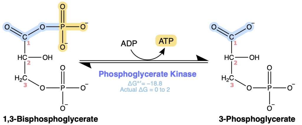
Figure X. Phosphoglycerate kinase transfers a phosphate group from 1,3-bisphosphoglycerate to ADP, generating ATP and 3-phosphoglycerate by substrate-level phosphorylation.
Mechanistic Description
PGK is essentially the reverse of PFK, but instead of using ATP to phosphorylate a sugar, it transfers the high-energy phosphate from 1,3-bisphosphoglycerate to ADP, producing the first ATP of glycolysis through substrate-level phosphorylation. The reaction begins when the terminal phosphate oxygen of ADP then acts as a nucleophile and attacks the electrophilic phosphorus atom of the C1 acyl phosphate group, while the electrons of the P=O bond shift onto the oxygen to form a pentavalent transition state. The electrons then collapse back to reform the P=O bond, while the electrons of the phosphate-oxygen bond shift onto the oxygen of 3-phosphoglycerate, releasing ATP and producing 3-phosphoglycerate. The defining feature of this mechanism is direct phosphoryl transfer through a pentavalent transition state. Unlike the ATP-consuming kinase reactions earlier in glycolysis, PGK transfers a high-energy phosphate from the substrate directly to ADP, producing the first ATP through substrate-level phosphorylation.
Just like the other kinases, PGK requires magnesium ions (Mg²⁺) for catalysis and to help stabilize the negatively charged phosphate groups of both ADP and ATP during the transition state.
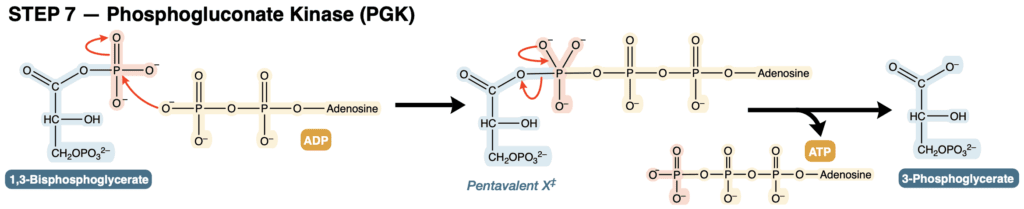
Figure X. Phosphoglycerate kinase transfers the high-energy phosphate from 1,3-bisphosphoglycerate to ADP, producing 3-phosphoglycerate and ATP through a pentavalent phosphoryl transition state.
Thermodynamics
The phosphoglycerate kinase reaction operates near equilibrium under physiological conditions.
ΔG°′ ≈ −18.8 kJ/mol
Although ATP synthesis is generally energetically unfavorable, this reaction proceeds because 1,3-bisphosphoglycerate (1,3-BPG) has an exceptionally high phosphoryl transfer potential. The free energy released by hydrolysis of its acyl phosphate bond is more than enough to drive ATP formation from ADP. Even though this reaction generates ATP, it is one of the few kinase reactions in metabolism that is readily reversible. Unlike most kinases, which catalyze reactions that strongly favor one direction, phosphoglycerate kinase (PGK) catalyzes a near-equilibrium reaction. As a result, it can readily operate in either direction depending on the concentrations of its substrates and products.
Regulation
Phosphoglycerate kinase is not considered a major regulatory enzyme of glycolysis.
Because the reaction operates close to equilibrium, its direction is determined primarily by substrate and product concentrations rather than extensive allosteric regulation. Glycolytic flux through this step is controlled largely by regulatory enzymes elsewhere in the pathway, particularly phosphofructokinase-1.
Other Notes
The End of an ATP Debt
For many students, this reaction marks a major milestone in glycolysis. The previous six reactions consumed ATP to prepare glucose for energy extraction, while PGK is the first enzyme that actually returns ATP to the cell. Earlier in the pathway, two ATP molecules were consumed by hexokinase and phosphofructokinase-1. Because PGK generates two ATP molecules per glucose, the pathway has now completely recovered its initial ATP investment. At this point in glycolysis, the net ATP yield is zero. Additional ATP generated later in the pathway will therefore represent a true energetic gain for the cell.
A Classic Example of Substrate-Level Phosphorylation
PGK provides one of the clearest examples of substrate-level phosphorylation in metabolism. Earlier, we discussed that ATP can be generated through either substrate-level phosphorylation or oxidative phosphorylation. In substrate-level phosphorylation, a phosphate group is transferred directly from a high-energy metabolic intermediate onto ADP. No electron transport chain, proton gradient, or ATP synthase is required. The phosphoglycerate kinase reaction is therefore fundamentally different from ATP production by oxidative phosphorylation, which will be discussed in a later post.
Figure X. text.
Coupling with the GAPDH Reaction
The GAPDH and phosphoglycerate kinase (PGK) reactions are tightly energetically coupled. GAPDH captures the energy released during oxidation by storing it in the high-energy acyl phosphate bond of 1,3-bisphosphoglycerate (1,3-BPG), and PGK immediately uses that stored energy to produce ATP through substrate-level phosphorylation. Because PGK rapidly consumes 1,3-BPG as it is formed, it helps prevent the intermediate from accumulating and continually pulls the GAPDH reaction forward. Together, these two reactions efficiently capture the energy released during oxidation and convert it into ATP.
Figure X. text.
The 2,3-Bisphosphoglycerate Shunt
In red blood cells, a portion of 1,3-BPG can bypass the phosphoglycerate kinase (PGK) reaction through the 2,3-bisphosphoglycerate (2,3-BPG) shunt. In this pathway, the bifunctional enzyme bisphosphoglycerate mutase (BPGM) first converts 1,3-BPG into 2,3-BPG through its mutase activity. The same enzyme then uses its phosphatase activity to hydrolyze 2,3-BPG into 3-phosphoglycerate. Because this bypasses the ATP-producing PGK reaction, no ATP is generated from that molecule of 1,3-BPG. Although this pathway reduces ATP production, it allows red blood cells to produce 2,3-BPG, which binds deoxyhemoglobin and decreases its affinity for oxygen. As a result, hemoglobin releases oxygen more readily to peripheral tissues.
Important Note: The 2,3-BPG shunt is most active in red blood cells, where 2,3-BPG reaches high concentrations to regulate oxygen delivery. However, small amounts of 2,3-BPG are also produced in many other cell types. In these cells, it functions in the activation of phosphoglycerate mutase (PGM), a topic that will be discussed in the following section.
Figure X. text.
STEP 8: Phosphoglycerate Mutase (PGM)
Reaction & Classifications
The eighth step of glycolysis is catalyzed by phosphoglycerate mutase (PGM), which converts 3-phosphoglycerate (3-PG) into 2-phosphoglycerate (2-PG). In this reaction, the phosphate group is moved from carbon 3 to carbon 2 of the molecule without changing the overall carbon skeleton.
Phosphoglycerate mutase belongs to the isomerase enzyme class because it catalyzes an intramolecular rearrangement. More specifically, it is classified as a mutase, a specialized type of isomerase that transfers a functional group from one position within a molecule to another.
Although no ATP is produced or consumed during this step, the reaction plays a critical role in preparing the molecule for the high-energy phosphate generation that occurs later in glycolysis.
Overall Reaction: 3-Phosphoglycerate ⇌ 2-Phosphoglycerate
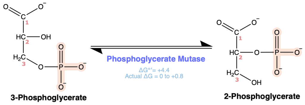
Figure X. Phosphoglycerate mutase reversibly transfers the phosphate group from C3 to C2, converting 3-phosphoglycerate to 2-phosphoglycerate.
Mechanistic Description
Phosphoglycerate mutase catalyzes an intramolecular phosphate shift by temporarily transferring a phosphate group to the substrate through a phosphohistidine intermediate. The reaction begins when an active site residue acts as a base to abstract the proton from the C2 hydroxyl group of 3-phosphoglycerate, allowing the resulting oxygen lone pair to attack the phosphorus atom of the phosphohistidine residue. As the new oxygen-phosphorus bond forms, the electrons of the P=O bond shift onto the oxygen, generating a pentavalent transition state. The electrons then collapse back to reform the P=O bond while the electrons of the phosphorus-nitrogen bond shift onto the histidine nitrogen, releasing a histidyl residue and producing the 2,3-bisphosphoglycerate intermediate. Next, the active site histidine acts as a nucleophile and attacks the phosphorus atom of the C3 phosphate group, while the electrons of the P=O bond shift onto the oxygen to form a second pentavalent transition state. The electrons then collapse back to reform the P=O bond as the electrons of the phosphate-oxygen bond shift onto the oxygen of the substrate, releasing the C3 phosphate group. Finally, the active site residue protonates the C3 oxygen, producing 2-phosphoglycerate and regenerating the phosphohistidine residue for another catalytic cycle. The defining feature of this mechanism is the transient formation of the 2,3-bisphosphoglycerate intermediate. By temporarily adding and then removing a phosphate group, phosphoglycerate mutase efficiently relocates the phosphate from C3 to C2 without requiring ATP. Also importantly, the enzyme is regenerated in its phosphorylated state at the end of the catalytic cycle, allowing it to continue catalysis without requiring ATP input for each reaction.

Figure X. Phosphoglycerate mutase transfers the phosphate group from C3 to C2 through a phosphohistidyl enzyme intermediate and the transient formation of 2,3-bisphosphoglycerate.
Thermodynamics
The phosphoglycerate mutase reaction operates close to equilibrium under physiological conditions.
ΔG°′ ≈ +4.4 kJ/mol
Although the standard free energy change is slightly positive, the actual cellular ΔG remains near zero because downstream reactions continuously consume 2-phosphoglycerate. Consequently, the reaction readily proceeds in either direction depending on metabolic conditions.
Regulation
Phosphoglycerate mutase is not considered a major regulatory enzyme of glycolysis.
The reaction operates near equilibrium, and its direction is determined primarily by substrate and product concentrations rather than allosteric regulation. As with many near-equilibrium glycolytic reactions, overall flux through this step is controlled largely by regulatory enzymes elsewhere in the pathway.
Other Notes
Why Move the Phosphate?
At first glance, moving a phosphate group from carbon 3 to carbon 2 may appear insignificant. However, this rearrangement is essential for the next stage of glycolysis. By repositioning the phosphate onto carbon 2, phosphoglycerate mutase prepares the substrate for the subsequent dehydration reaction catalyzed by enolase, which generates phosphoenolpyruvate (PEP), one of the highest-energy phosphate-containing intermediates in metabolism. If the phosphate remained on carbon 3, dehydration would produce a different molecule rather than PEP, preventing the formation of the high-energy phosphate donor required for ATP production by pyruvate kinase.
Figure X. text.
An Alternative Role of 2,3-BPG
Although the highest concentrations of 2,3-BPG are found in red blood cells, small amounts are also produced in many other tissues. This is because PGM contains an active-site histidine residue that must remain phosphorylated for the enzyme to function. Occasionally, this phosphohistidine is lost through hydrolysis, leaving the enzyme inactive. Small amounts of 2,3-BPG act as a phosphate donor to rephosphorylate the histidine residue and restore PGM activity. The 2,3-BPG that reactivates PGM is not the same 2,3-BPG intermediate formed during the PGM reaction. Instead, it serves only to regenerate the phosphorylated enzyme before catalysis begins. This maintenance role explains why many tissues produce low levels of 2,3-BPG even though they do not use it to regulate hemoglobin oxygen affinity.
STEP 9: Enolase
Reaction & Classifications
The ninth step of glycolysis is catalyzed by enolase, which converts 2-phosphoglycerate (2-PG) into phosphoenolpyruvate (PEP). This reaction removes a molecule of water from the substrate, generating a carbon-carbon double bond and producing an enol phosphate.
Enolase belongs to the lyase enzyme class because it removes a group from a substrate without hydrolysis or oxidation. More specifically, the reaction is classified as a dehydration reaction.
Although no ATP is produced during this step, the reaction generates phosphoenolpyruvate (PEP), which possesses one of the highest phosphoryl transfer potentials found in metabolism. This prepares the substrate for the final ATP-generating step of glycolysis.
Overall Reaction: 2-Phosphoglycerate ⇌ Phosphoenolpyruvate + H₂O
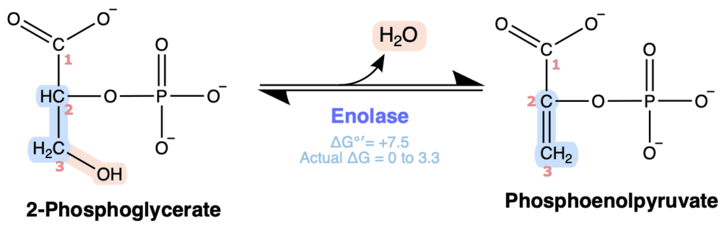
Figure X. Enolase catalyzes the reversible dehydration of 2-phosphoglycerate to form phosphoenolpyruvate, releasing one molecule of water.
Mechanistic Description
Enolase catalyzes a dehydration reaction that removes water from 2-phosphoglycerate, generating the high-energy enol phosphate phosphoenolpyruvate (PEP). The reaction begins when an active site residue acts as a base to abstract the proton from C2 of 2-phosphoglycerate. The resulting electrons form a C2=C3 double bond as the C3 hydroxyl group is protonated, converting it into a better leaving group. The electrons of the C3-O bond then shift onto the oxygen, releasing water and producing phosphoenolpyruvate. The defining feature of this mechanism is β-elimination of water to form the high-energy enol phosphate bond of phosphoenolpyruvate. Formation of this enol phosphate stores substantial free energy within the product, enabling phosphoenolpyruvate to donate its phosphate group to ADP during the next step of glycolysis.
Two Mg²⁺ ions participate in catalysis and help stabilize negatively charged intermediates that develop during the reaction.
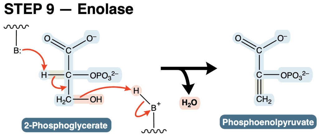
Figure X. Enolase catalyzes the dehydration of 2-phosphoglycerate to phosphoenolpyruvate by eliminating water and forming a high-energy enol phosphate.
Thermodynamics
The enolase reaction has a slightly positive standard free energy change.
ΔG°′ ≈ +1.8 kJ/mol
Despite this value, the reaction operates close to equilibrium under cellular conditions because phosphoenolpyruvate is rapidly consumed by pyruvate kinase in the next step of glycolysis.
Regulation
Enolase is not considered a major regulatory enzyme of glycolysis. The reaction operates near equilibrium, and its direction is determined primarily by substrate and product concentrations. Overall glycolytic flux through this step is therefore controlled largely by regulatory enzymes elsewhere in the pathway.
Other Notes
Mechanistic Importance
The physiological importance of this reaction lies in the product rather than the reaction itself. Enolase converts a relatively low-energy phosphoglycerate into phosphoenolpyruvate, one of the highest-energy phosphate-containing compounds in metabolism. At first glance, it may seem counterintuitive that simply removing water creates such a high-energy molecule. However, the dehydration reaction generates an unstable enol phosphate. Hydrolysis of PEP is highly favorable because the resulting enol product rapidly undergoes tautomerization to the much more stable keto form of pyruvate. This additional stabilization greatly increases the free energy released during hydrolysis. As a result, PEP possesses a greater phosphoryl transfer potential than ATP itself.
Figure X. text.
Note
text
Figure X. text.
STEP 10: Pyruvate Kinase (PK)
Reaction & Classifications
The tenth and final step of glycolysis is catalyzed by pyruvate kinase, which transfers a phosphate group from phosphoenolpyruvate (PEP) to ADP, producing pyruvate and ATP.
Pyruvate kinase belongs to the transferase enzyme class, specifically the kinase subclass, because it transfers a phosphate group from one molecule to another. Like phosphoglycerate kinase, the reaction is classified as a substrate-level phosphorylation because ATP is generated directly through phosphate transfer rather than through oxidative phosphorylation.
This reaction represents the second ATP-producing step of glycolysis. Because two molecules of phosphoenolpyruvate are generated from each glucose molecule, the reaction occurs twice per glucose and produces two ATP molecules. These two ATP molecules constitute the net ATP gain of glycolysis because the pathway previously consumed two ATP molecules during the energy investment phase.
Overall Reaction: Phosphoenolpyruvate + ADP → Pyruvate + ATP
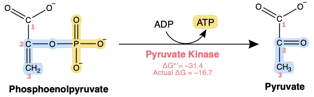
Figure X. Pyruvate kinase transfers the phosphate group from phosphoenolpyruvate to ADP, generating ATP and pyruvate by substrate-level phosphorylation.
Mechanistic Description
Pyruvate kinase catalyzes the second substrate-level phosphorylation of glycolysis by transferring the high-energy phosphate group from phosphoenolpyruvate (PEP) to ADP, producing ATP. The reaction begins when the terminal phosphate oxygen of ADP acts as a nucleophile and attacks the electrophilic phosphorus atom of phosphoenolpyruvate, while the electrons of the P=O bond shift onto the oxygen to form a pentavalent transition state. The electrons then collapse back to reform the P=O bond, while the electrons of the phosphate-oxygen bond shift onto the oxygen of phosphoenolpyruvate, transferring the phosphate group to ADP and producing ATP. Loss of the phosphate group generates the unstable enol form of pyruvate, which rapidly undergoes keto-enol tautomerization as the electrons of the C=C bond shift to reform the carbonyl and C3 is protonated, producing the more stable keto form of pyruvate. The defining feature of this mechanism is coupling substrate-level phosphorylation with spontaneous keto-enol tautomerization of pyruvate. Formation of the thermodynamically favored keto form drives the reaction strongly forward, making pyruvate kinase one of the three essentially irreversible steps of glycolysis.
Mg²⁺ and K⁺ ions are required cofactors for catalysis. These ions help stabilize negative charges that develop during phosphoryl transfer and properly orient the substrates within the active site.
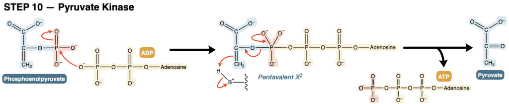
Figure X. Pyruvate kinase transfers the phosphate group from phosphoenolpyruvate to ADP, producing ATP and pyruvate through a pentavalent phosphoryl transition state followed by enol-to-keto tautomerization.
Thermodynamics
The pyruvate kinase reaction is strongly exergonic and essentially irreversible under physiological conditions.
ΔG°′ ≈ −31.4 kJ/mol
The large negative free energy change arises largely from the exceptionally high phosphoryl transfer potential of phosphoenolpyruvate and the subsequent stabilization gained through conversion of enol-pyruvate to pyruvate. As discussed in the previous section, phosphoenolpyruvate is one of the highest-energy phosphate-containing intermediates found in metabolism. Hydrolysis of its phosphate group is highly favorable, and the additional stabilization provided by keto-enol tautomerization makes the overall reaction strongly irreversible.
Regulation
regulate
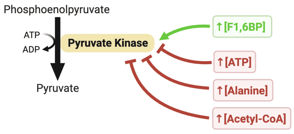
Figure X. Regulation of pyruvate kinase. Fructose-1,6-bisphosphate feed-forward activates the enzyme, whereas ATP, alanine, and acetyl-CoA inhibit its activity, allowing glycolytic flux to respond to cellular energy status and biosynthetic needs.
Other Notes
From a physiological perspective, pyruvate kinase completes the extraction of usable energy from glycolysis. The ATP generated during this reaction represents the pathway’s net ATP yield, and the pyruvate produced serves as an important metabolic branch point that can enter numerous downstream pathways depending on cellular conditions.
Under aerobic conditions, pyruvate is typically converted to acetyl-CoA and enters the citric acid cycle. Under anaerobic conditions, pyruvate can instead undergo fermentation reactions that regenerate NAD⁺ and allow glycolysis to continue.
Figure X. Regulation of pyruvate kinase. Fructose-1,6-bisphosphate feed-forward activates the enzyme, whereas ATP, alanine, and acetyl-CoA inhibit its activity, allowing glycolytic flux to respond to cellular energy status and biosynthetic needs.