
Glucose Digestion, Absorption, Transport, & Uptake
Introduction to Why Glucose Matters
Glucose is one of the most important molecules in metabolism. Nearly every cell uses glucose either as an energy source or building block for the synthesis of other molecules. Cells break down glucose through glycolysis, the citric acid cycle, and oxidative phosphorylation to produce ATP, the cell’s primary energy currency. Glucose also supplies carbon for the synthesis of glycogen, nucleotides, amino acids, lipids, and many other biomolecules.
Although some tissues can utilize alternative fuels (like fats) under certain conditions, others depend heavily on a continuous glucose supply. Red blood cells rely entirely on glucose because they lack mitochondria, while the brain consumes large amounts of glucose under normal physiological conditions. Maintaining blood glucose concentrations within a narrow range is therefore essential for normal cellular and organ function.
Before glucose can participate in metabolism, the body must first obtain carbohydrates from the diet. The digestive system breaks these carbohydrates into absorbable sugars, transports them across the intestinal epithelium, and delivers them to the bloodstream. From there, glucose travels to tissues throughout the body, where cells take it up and direct it into metabolic pathways. Understanding this journey provides an important foundation for understanding how glucose enters pathways such as glycolysis.
Dietary Sources of Glucose
Dietary carbohydrates provide most of the glucose used by the body. Although these carbohydrates vary considerably in size and structure, digestion ultimately reduces them to monosaccharides that can be absorbed by the intestine and transported to tissues throughout the body.
The simplest carbohydrates are monosaccharides, which consist of a single sugar unit. The major dietary monosaccharides are glucose, fructose, galactose, and mannose. Each contains six carbon atoms but differs in the arrangement of functional groups around the carbon skeleton. Glucose, galactose, and mannose are aldohexoses that contain an aldehyde group at carbon 1 in their open-chain forms and typically cyclize into 6-membered pyranose rings. Fructose, in contrast, is a ketohexose with a ketone group at carbon 2 and most commonly cyclizes into a 5-membered furanose ring, although pyranose forms also exist. Monosaccharides are the final products of carbohydrate digestion and the forms of carbohydrate that can be absorbed by the intestinal epithelium.
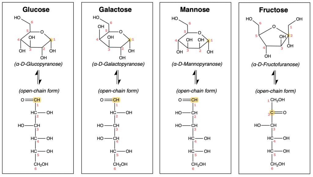
Figure 2. Structures of the major dietary monosaccharides. Glucose, galactose, and mannose are aldohexoses that typically cyclize into six-membered pyranose rings, whereas fructose is a ketohexose that commonly forms a five-membered furanose ring.
Many foods also contain disaccharides, which consist of two monosaccharides joined by a glycosidic bond. The three common dietary disaccharides are sucrose, lactose, and maltose. Sucrose consists of glucose and fructose linked through an α(1→2) glycosidic bond between the anomeric carbons of both sugars. It is abundant in fruits, table sugar, and many sweetened foods. Lactose consists of galactose and glucose joined by a β(1→4) glycosidic bond. It is the major carbohydrate in milk and other dairy products. Maltose consists of two glucose molecules connected by an α(1→4) glycosidic bond. Although maltose is present in malted grains and some fermented foods, it is most commonly produced during starch digestion. Because the intestine can absorb only monosaccharides, these disaccharides must first be hydrolyzed into their individual sugar components.
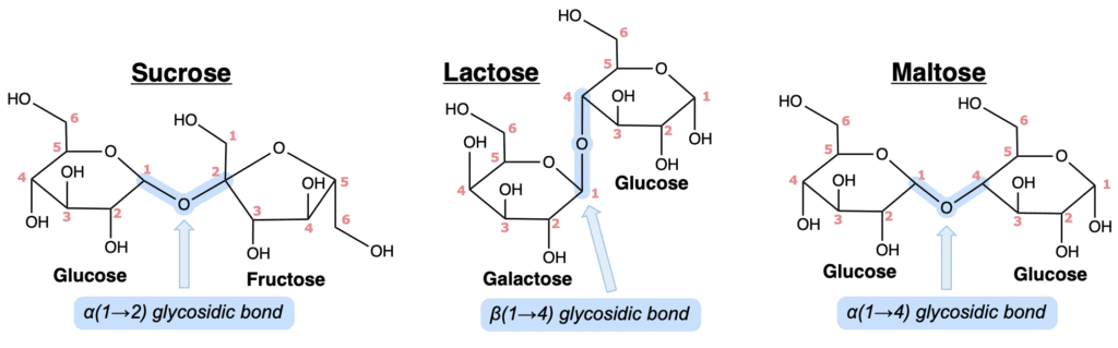
Figure 3. Structures of the major dietary disaccharides. Sucrose contains an α(1→2) linkage between glucose and fructose, lactose contains a β(1→4) linkage between galactose and glucose, and maltose contains an α(1→4) linkage between two glucose residues.
Most dietary carbohydrates are consumed as polysaccharides, which are long polymers composed primarily of glucose residues. The most abundant dietary polysaccharide is starch, the primary storage carbohydrate of plants. Starch exists as two major forms: amylose and amylopectin. Amylose is a largely linear polymer of glucose residues connected by α(1→4) glycosidic bonds. Amylopectin contains the same α(1→4)-linked glucose backbone but also possesses α(1→6) branch points approximately every 24 to 30 glucose residues. Starch provides most of the carbohydrate in the human diet and is abundant in grains, bread, pasta, cereals, potatoes, and other starchy plant foods.
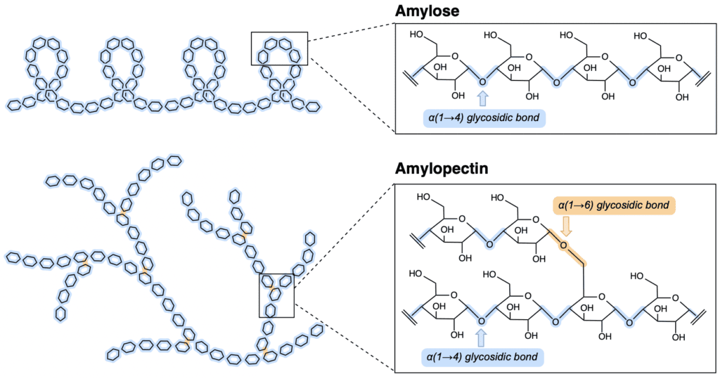
Figure 4. The two major forms of dietary starch. Amylose is a largely linear glucose polymer linked by α(1→4) glycosidic bonds, whereas amylopectin contains the same α(1→4)-linked backbone with additional α(1→6) branch points.
Glycogen, the primary storage form of glucose in animals, possesses a structure similar to amylopectin but is more extensively branched. Similar to amylopectin, glycogen contains α(1→4) glycosidic bonds along its linear chains and α(1→6) glycosidic bonds at branch points. However, glycogen branches approximately every 8 to 12 glucose residues, producing a highly compact structure that allows rapid glucose mobilization when needed.
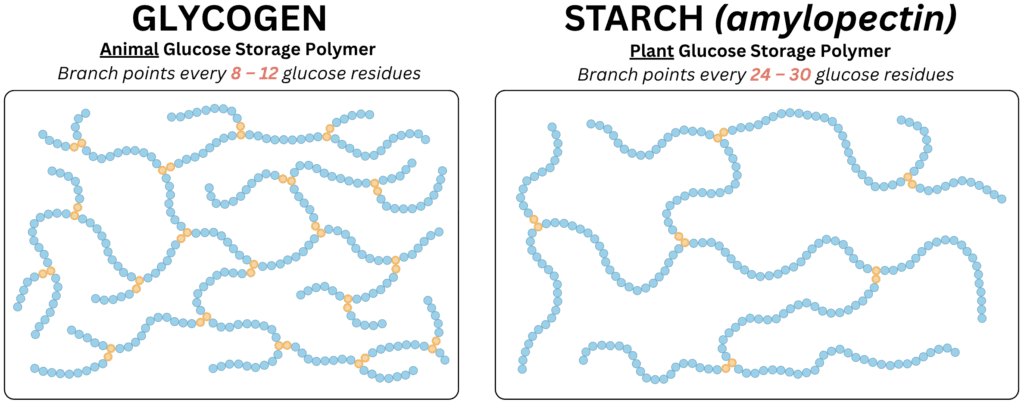
Figure 5. Comparison of the two major glucose storage polysaccharides. Glycogen, the storage form of glucose in animals, contains α(1→6) branch points approximately every 8 to 12 glucose residues, whereas amylopectin, the branched form of starch in plants, contains branch points approximately every 24 to 30 glucose residues.
In contrast, dietary fiber consists largely of polysaccharides that humans cannot efficiently digest. The most abundant example is cellulose, a linear polymer of glucose residues connected by β(1→4) glycosidic bonds. Although cellulose is composed entirely of glucose, humans lack cellulase, the enzyme required to hydrolyze β(1→4) linkages. As a result, cellulose passes through the digestive tract largely undigested.
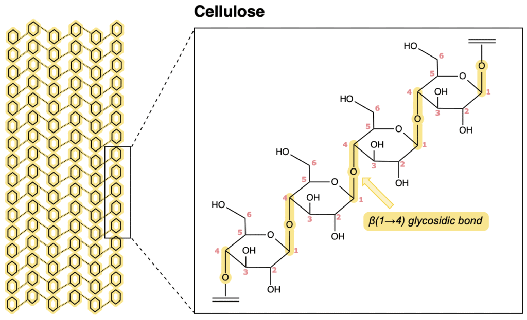
Figure 6. Structure of cellulose, the primary structural polysaccharide of plants. Glucose residues are connected by β(1→4) glycosidic bonds, producing straight chains that assemble into strong fibers resistant to digestion by human enzymes.
Carbohydrate Digestion
Carbohydrate digestion occurs through a series of mechanical and enzymatic processes that progressively break large carbohydrate molecules into absorbable monosaccharides.
Digestion begins in the mouth. Chewing mechanically fragments food into smaller particles, increasing the surface area available for enzymatic activity. At the same time, salivary α-amylase initiates carbohydrate digestion by hydrolyzing internal α(1→4) glycosidic bonds within starch and glycogen. Because α-amylase cleaves only internal α(1→4) linkages, it cannot hydrolyze terminal glucose residues, α(1→6) branch points, disaccharides, or trisaccharides. The products of salivary digestion therefore consist primarily of maltose, maltotriose, short oligosaccharides, α-limit dextrins (short branch points), and other incompletely digested starch molecules. These α-limit dextrins contain the α(1→6) branch points that α-amylase cannot hydrolyze.
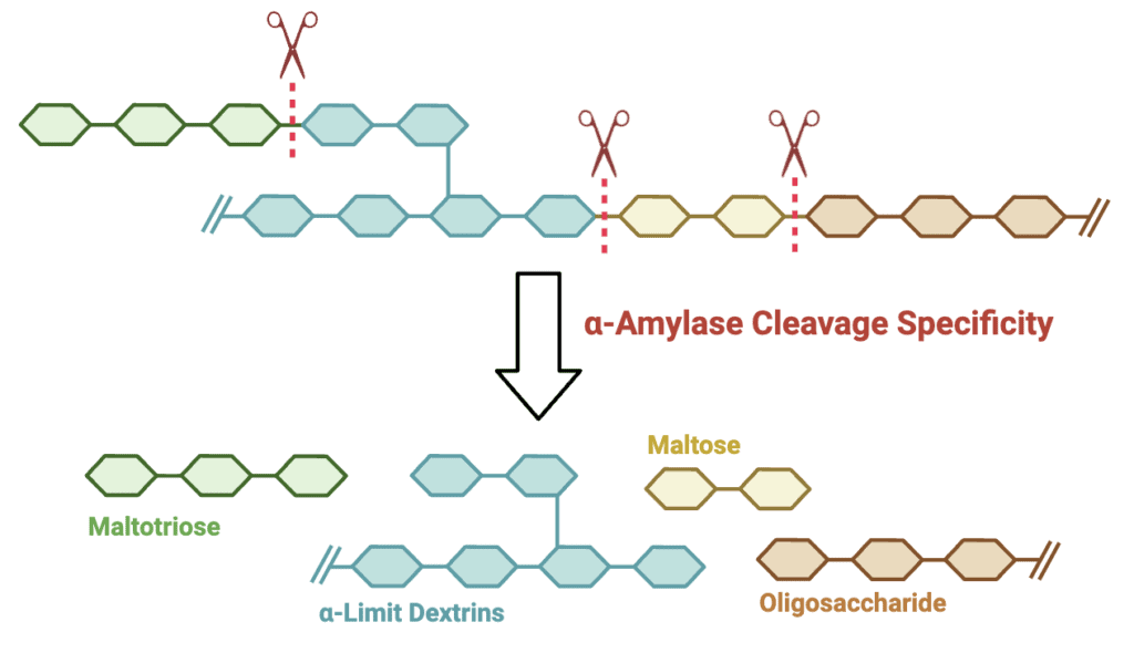
Figure 7. α-Amylase hydrolyzes internal α(1→4) glycosidic bonds within starch, generating smaller carbohydrates such as maltose, maltotriose, oligosaccharides, and α-limit dextrins while leaving α(1→6) branch points intact.
After swallowing, digestion temporarily slows in the stomach. The acidic gastric environment gradually inactivates salivary α-amylase, and little additional enzymatic digestion of carbohydrates occurs. The primary contribution of the stomach is mechanical mixing, which disperses food and delivers it to the small intestine.
Most carbohydrate digestion occurs in the small intestine. Pancreatic α-amylase, secreted into the intestinal lumen by the pancreas, continues hydrolyzing internal α(1→4) glycosidic bonds within starch and glycogen. Because it possesses the same substrate specificity as salivary α-amylase, pancreatic α-amylase also produces maltose, maltotriose, α-limit dextrins, and some oligosaccharides. Although the same bonds are cleaved, the greater extent of digestion in the small intestine allows large starch and glycogen polymers that escaped salivary digestion to be further degraded into smaller carbohydrates.
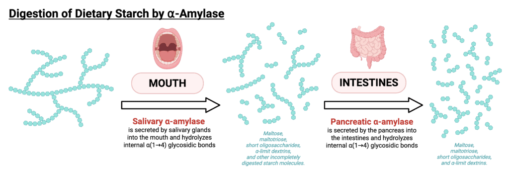
Figure 8. Starch digestion begins in the mouth with salivary α-amylase and continues in the small intestine with pancreatic α-amylase. Both enzymes hydrolyze internal α(1→4) glycosidic bonds, producing maltose, maltotriose, oligosaccharides, and α-limit dextrins.
The final stages of digestion occur at the brush border of the intestinal epithelium. Several membrane-associated enzymes hydrolyze the remaining carbohydrates into absorbable monosaccharides. Unlike the α-amylases, which are endoglycosidases that cleave internal glycosidic bonds, brush-border enzymes function as exoglycosidases, hydrolyzing terminal glycosidic bonds to release individual monosaccharides.
Two brush-border enzymes complete the digestion of starch-derived carbohydrates. Maltase hydrolyzes terminal α(1→4) glycosidic bonds in maltose and short oligosaccharides, producing glucose. Isomaltase hydrolyzes α(1→6) glycosidic bonds at branch points within α-limit dextrins, allowing complete digestion of branched starch and glycogen fragments into glucose.
Although starch is the predominant carbohydrate in most diets, other dietary disaccharides also require specialized brush-border enzymes. Sucrase hydrolyzes the α(1→2) glycosidic bond of sucrose, releasing glucose and fructose. Lactase hydrolyzes the β(1→4) glycosidic bond of lactose, producing glucose and galactose.
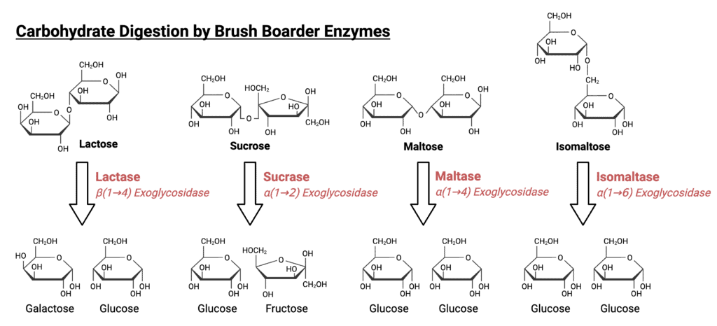
Figure 9. Brush-border enzymes complete carbohydrate digestion by hydrolyzing disaccharides and oligosaccharides into absorbable monosaccharides. Lactase, sucrase, maltase, and isomaltase cleave specific glycosidic bonds to generate glucose, galactose, and fructose.
The overall process of carbohydrate digestion can therefore be summarized as follows:
- Salivary α-amylase begins hydrolysis of internal α(1→4) glycosidic bonds.
- Pancreatic α-amylase continues hydrolysis of internal α(1→4) glycosidic bonds.
- Maltase hydrolyzes maltose and short α(1→4)-linked oligosaccharides.
- Isomaltase hydrolyzes α(1→6) branch points.
- Sucrase hydrolyzes sucrose.
- Lactase hydrolyzes lactose.
By the end of digestion, most dietary carbohydrates have been reduced to glucose, fructose, and galactose. These monosaccharides are now ready to cross the intestinal epithelium and enter the body.
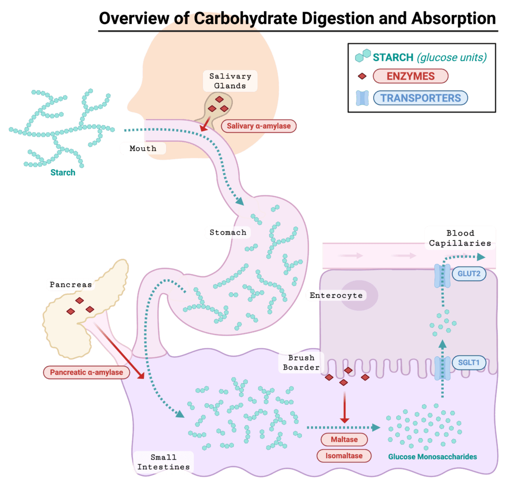
Figure 10. Overview of starch digestion and glucose absorption. Salivary and pancreatic α-amylases progressively hydrolyze starch into smaller carbohydrates, which are further digested by brush-border enzymes into monosaccharides that are absorbed by enterocytes and transported into the bloodstream.
Intestinal Absorption of Monosaccharides
Once digestion is complete, monosaccharides must move from the intestinal lumen into the bloodstream. This step takes place primarily in the small intestine and relies on specialized transport proteins located on the apical (facing the intestinal lumen) and basolateral (facing the bloodstream) membranes of enterocytes.
On the apical membrane, glucose and galactose enter enterocytes through the sodium-glucose cotransporter SGLT1. This transporter uses secondary active transport, coupling monosaccharide uptake to the movement of sodium ions into the cell. Because sodium moves down its electrochemical gradient, SGLT1 can transport glucose and galactose into the enterocyte even when intracellular concentrations are already relatively high.
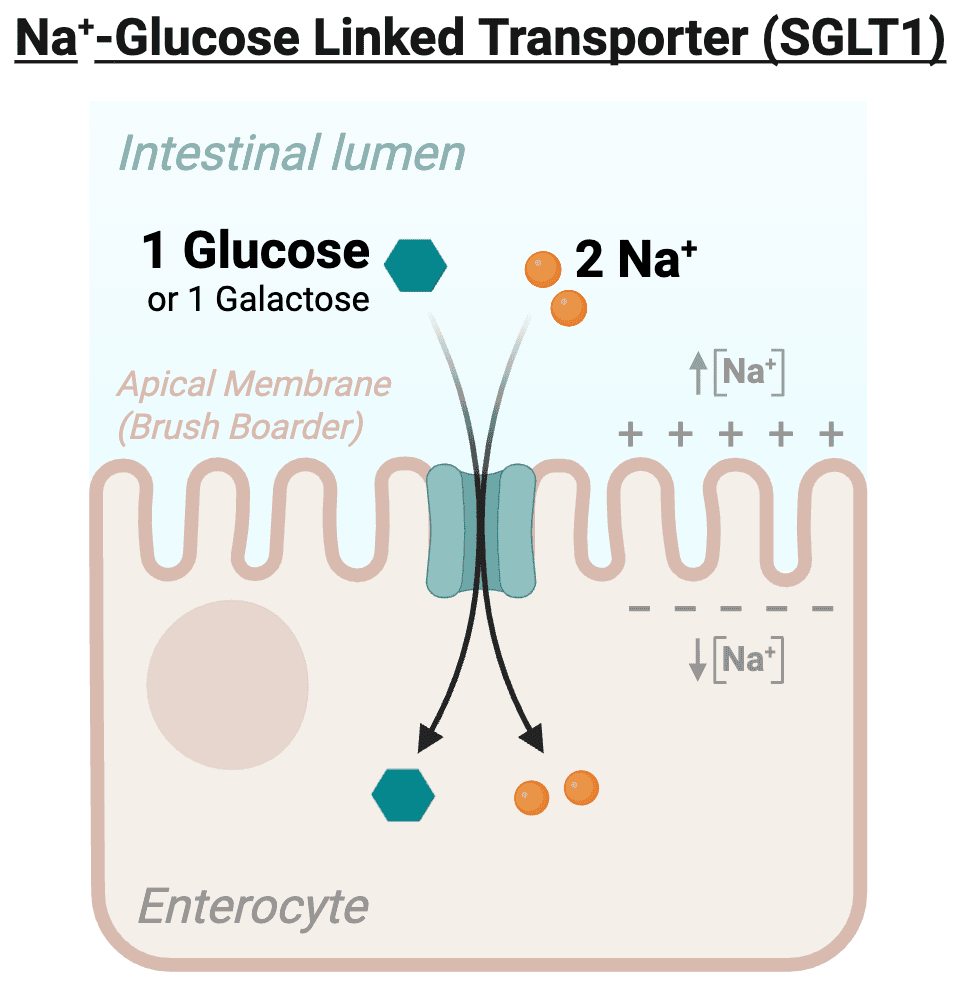
Figure 11. SGLT1 mediates uptake of glucose and galactose across the apical membrane of enterocytes by coupling monosaccharide transport to the inward movement of sodium ions down their electrochemical gradient.
The Na⁺/K⁺-ATPase on the basolateral membrane maintains the sodium gradient that powers SGLT1. By using ATP to pump sodium out of the cell and potassium into the cell, this transporter keeps intracellular sodium concentrations low. This electrochemical gradient stores potential energy that drives glucose and galactose uptake.
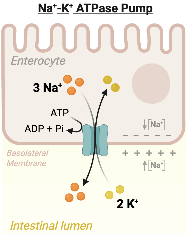
Figure 12. The basolateral Na⁺/K⁺-ATPase uses ATP to export three Na⁺ ions and import two K⁺ ions, maintaining the sodium gradient that drives secondary active transport of glucose and galactose by SGLT1.
Fructose follows a different route. Rather than relying on sodium-coupled transport, fructose enters enterocytes through GLUT5, a facilitative transporter that allows movement down its concentration gradient.
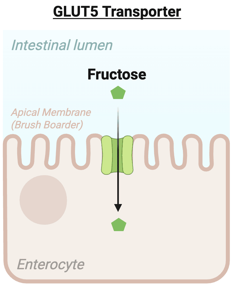
Figure 13. GLUT5 transports fructose across the apical membrane of enterocytes by facilitated diffusion, allowing fructose to move down its concentration gradient from the intestinal lumen into the cell.
After entering the enterocyte, all three monosaccharides exit across the basolateral membrane through GLUT2. They then enter the hepatic portal circulation, which delivers absorbed nutrients directly to the liver. The liver determines much of the metabolic fate of these sugars by storing, utilizing, or converting them into other molecules.
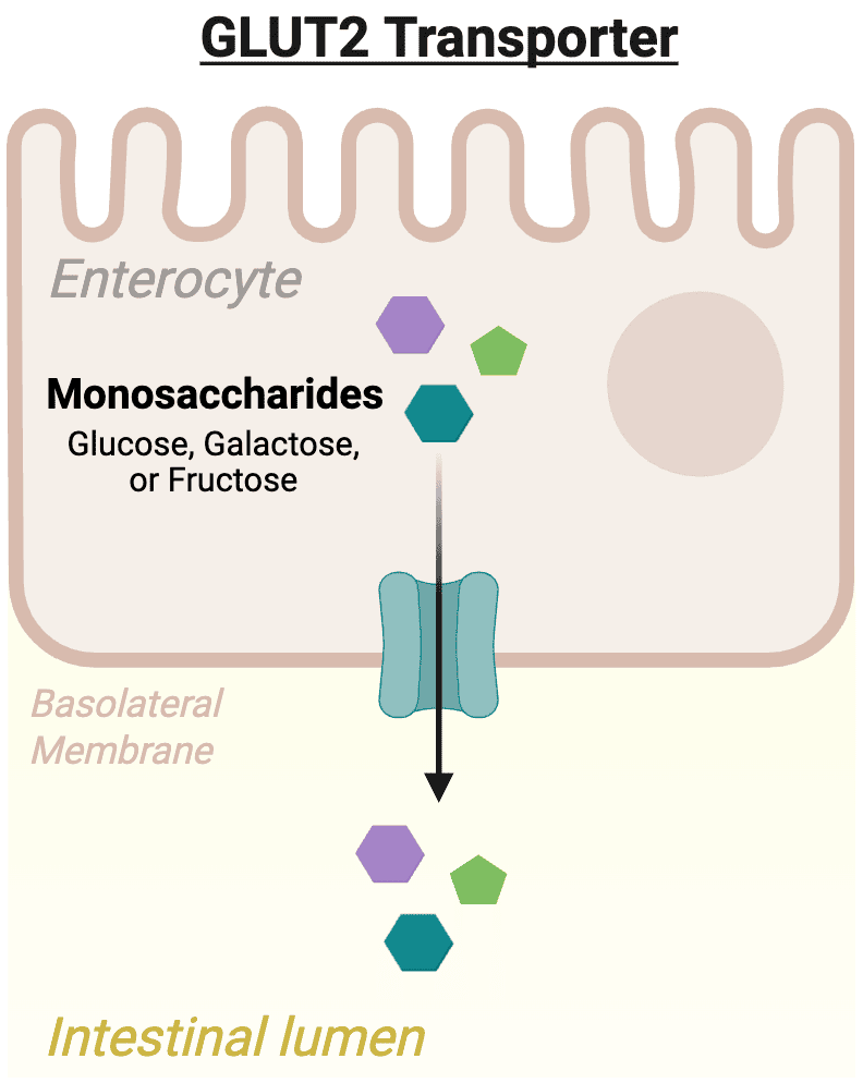
Figure 14. GLUT2 facilitates the export of glucose, galactose, and fructose across the basolateral membrane of enterocytes into the portal circulation by facilitated diffusion.
Below is a diagram summarizing the absorption of dietary carbohydrates from the intestines into the bloodstream.
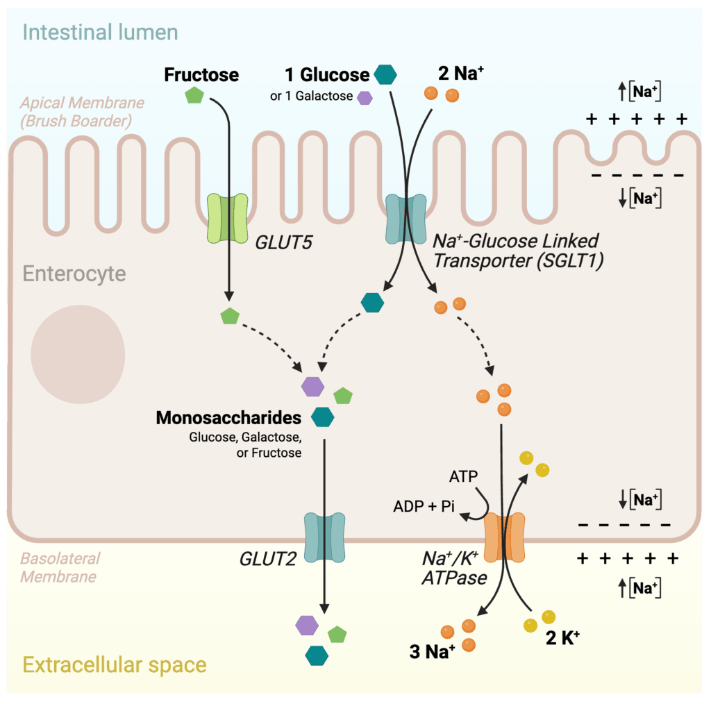
Figure 15. Absorption of dietary monosaccharides across the intestinal epithelium. Glucose and galactose enter enterocytes through SGLT1 by Na⁺-coupled secondary active transport, whereas fructose enters through GLUT5 by facilitated diffusion. All three monosaccharides exit the cell through GLUT2 on the basolateral membrane.
At this point, glucose has completed digestion and absorption. The next challenge is distributing glucose throughout the body and regulating its uptake into individual cells according to their metabolic needs.
Hepatic Processing of Absorbed Glucose
After absorption, glucose enters the hepatic portal circulation and is transported directly to the liver through the portal vein. This arrangement allows the liver to process incoming nutrients before they reach the rest of the body. As a result, the liver serves as an important metabolic buffer, helping prevent large fluctuations in blood glucose concentration following a meal.
Hepatocytes express GLUT2, a glucose transporter with relatively low affinity but high transport capacity. These kinetic properties allow the liver to respond efficiently to changes in blood glucose concentration. When glucose levels rise after a meal, GLUT2 facilitates rapid uptake into hepatocytes. When glucose levels fall during fasting, GLUT2 can also mediate glucose export from the liver. This bidirectional transport enables the liver to play a central role in maintaining blood glucose homeostasis.
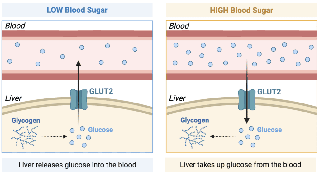
Figure 16. GLUT2 enables bidirectional glucose transport across the hepatocyte plasma membrane. During fasting, the liver releases glucose into the bloodstream, whereas after a meal it takes up glucose for storage and metabolism.
Once glucose enters hepatocytes, it can follow several different fates depending on the nutritional state of the body. Some glucose is used immediately for energy production, some is stored for later use, and some may be converted into other biologically important molecules. The specific pathways involved will be discussed in greater detail in future posts, but the key concept is that the liver helps determine how absorbed glucose is distributed and utilized throughout the body. Since all absorbed monosaccharides pass through the liver before reaching the systemic circulation, the liver is uniquely positioned to regulate the processing and distribution of dietary carbohydrates.
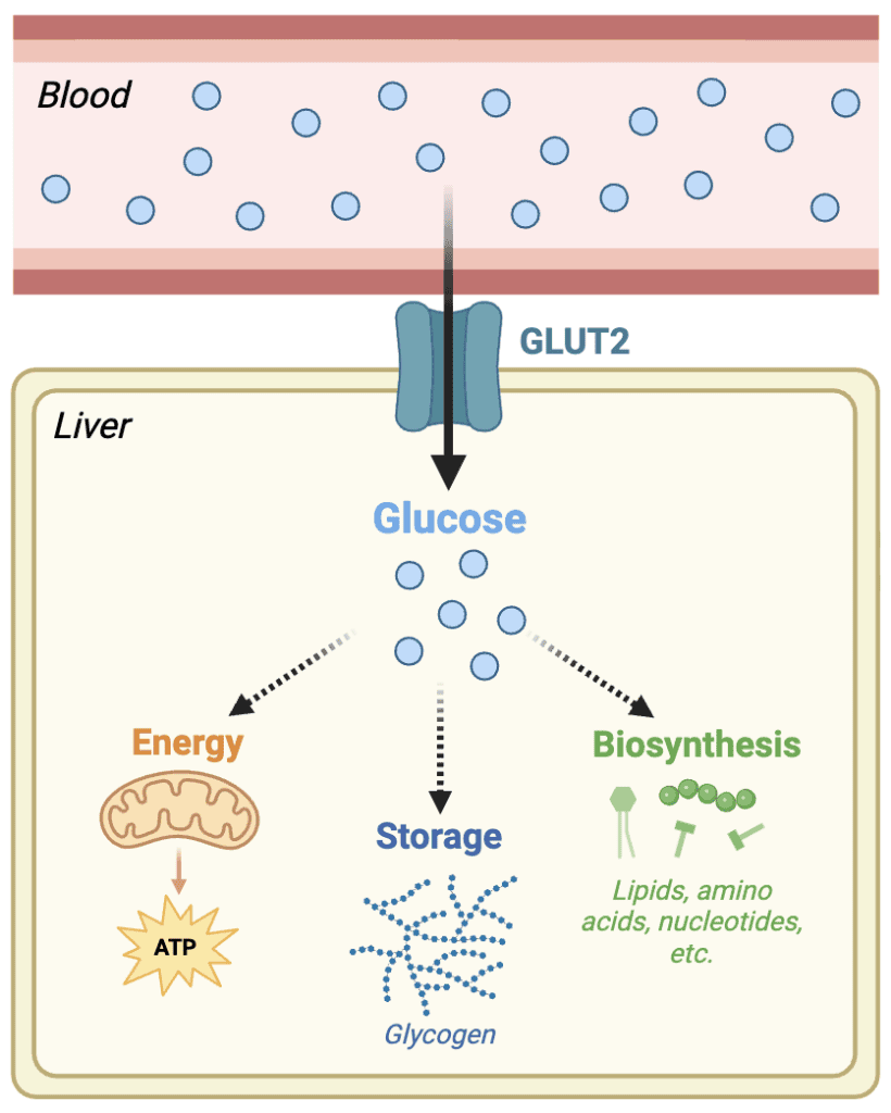
Figure 17. After entering hepatocytes through GLUT2, glucose can be oxidized to produce ATP, stored as glycogen, or used as a precursor for the synthesis of lipids, amino acids, nucleotides, and other biomolecules.
Blood Glucose Homeostasis
Despite continual changes in nutrient intake and energy expenditure, blood glucose concentration is maintained within a relatively narrow range. Under normal fasting conditions, blood glucose concentrations are approximately 5 mM (about 90 mg/dL). Maintaining glucose within this range is essential because both excessively low and excessively high blood glucose concentrations can impair normal cellular function.
Following a carbohydrate-containing meal, glucose absorbed from the intestine enters the bloodstream and causes blood glucose concentrations to rise. In response, pancreatic β-cells secrete insulin. Insulin promotes glucose uptake by insulin-responsive tissues and encourages storage of excess nutrients, helping return blood glucose concentrations toward their normal range.
During fasting, exercise, or periods between meals, blood glucose concentrations begin to decline. In response, pancreatic α-cells secrete glucagon. Glucagon stimulates the release of stored glucose and promotes mechanisms that help maintain an adequate supply of glucose for tissues that depend heavily on it.
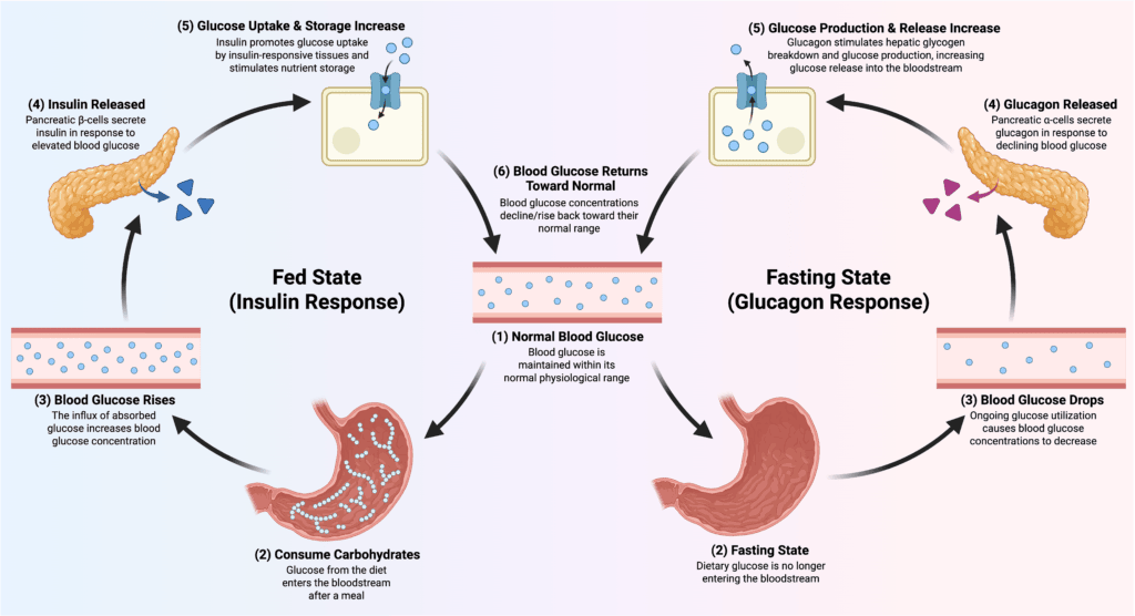
Figure 18. Blood glucose homeostasis is maintained through the opposing actions of insulin and glucagon. Following a carbohydrate-rich meal, insulin promotes glucose uptake and storage, whereas fasting stimulates glucagon release, increasing hepatic glucose production and glucose release to restore normal blood glucose concentrations.
Together, insulin and glucagon function as opposing hormonal regulators that help stabilize blood glucose concentrations despite continual changes in nutrient availability. Rather than allowing large fluctuations after every meal or prolonged fast, these hormones coordinate nutrient storage and mobilization to maintain a relatively constant supply of glucose throughout the body. This regulation is particularly important for tissues such as the brain and red blood cells, which rely heavily on glucose metabolism for normal function. By maintaining a stable supply of glucose, the body ensures that these tissues continue to receive the fuel needed to support cellular activity.
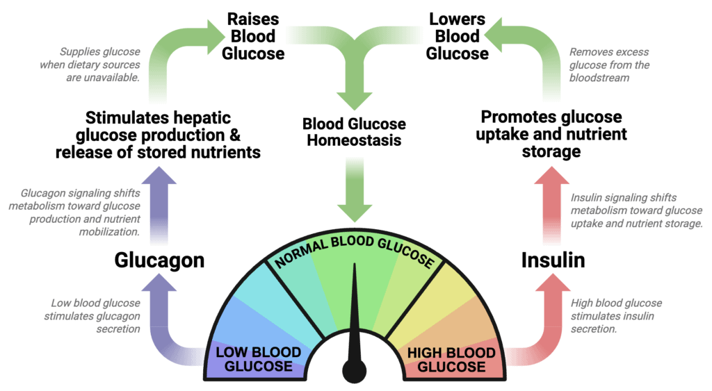
Figure 19. Insulin and glucagon work together through opposing negative feedback mechanisms to maintain blood glucose homeostasis. Insulin lowers blood glucose by promoting glucose uptake and storage, whereas glucagon raises blood glucose by stimulating hepatic glucose production and nutrient mobilization.
Glucose Transporters
After glucose enters the bloodstream, it must cross the plasma membrane to enter individual cells. Because glucose is a polar molecule, it cannot readily diffuse through the hydrophobic lipid bilayer. Instead, most tissues rely on a family of membrane transport proteins known as glucose transporters (GLUTs).

Figure 20. Crystal structure of the human GLUT3 glucose transporter in the outward-open conformation (PDB: 4ZWC; Deng et al., 2015).
Unlike the sodium-dependent transporters involved in intestinal absorption, GLUT proteins mediate facilitated diffusion. In facilitated diffusion, glucose moves down its concentration gradient without direct ATP consumption. The transporter simply provides a pathway that allows glucose to cross the membrane more efficiently. Different tissues express different GLUT isoforms, allowing glucose uptake to be customized to the metabolic needs of each tissue.
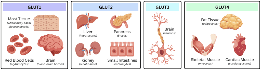
Figure 21. Major tissue distribution of the GLUT transporter family. GLUT1 supports basal glucose uptake in most tissues, GLUT2 is expressed in the liver, pancreatic β-cells, kidney, and enterocytes, GLUT3 is the primary neuronal glucose transporter, and GLUT4 mediates insulin-responsive glucose uptake in skeletal muscle, cardiac muscle, and adipose tissue.
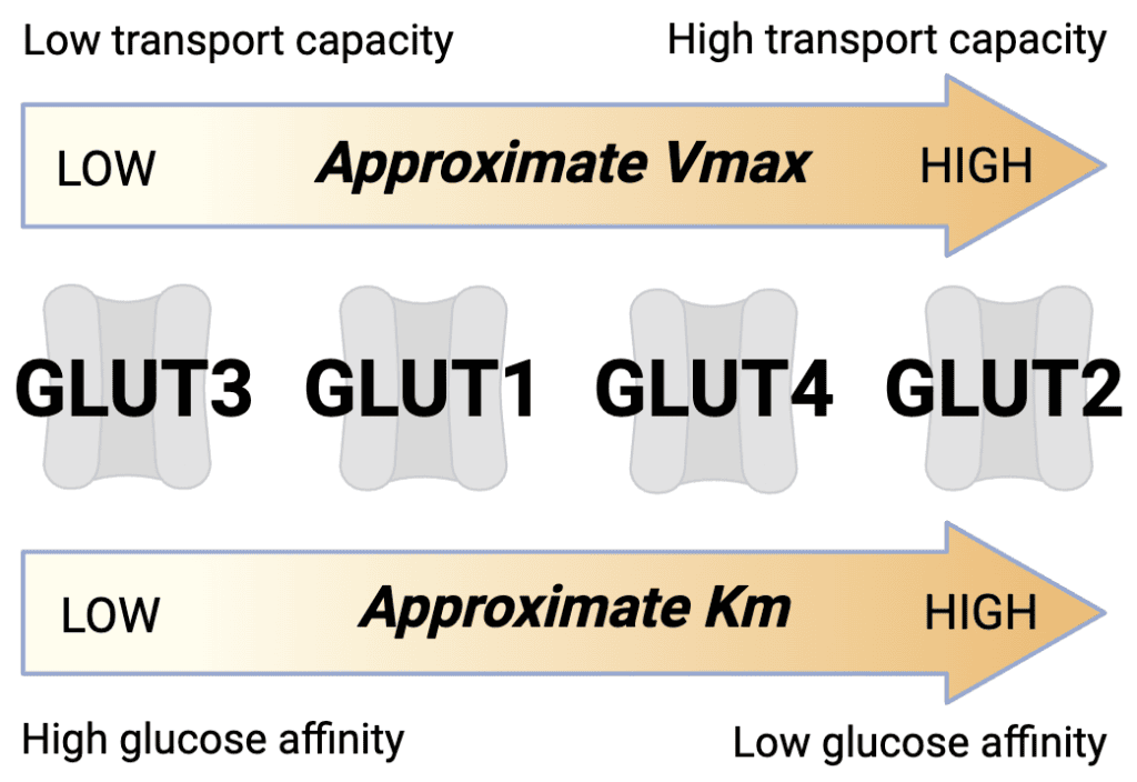
GLUT1 is widely distributed throughout the body for basal glucose uptake and is especially important in red blood cells and the blood-brain barrier. It has a high affinity for glucose, allowing efficient uptake even when blood glucose concentrations are relatively low. This helps ensure a continuous supply of glucose to tissues who heavily rely on uninterrupted energy availability.
GLUT2 is found primarily in the liver, pancreatic β-cells, small intestine, and kidney. Compared to the other transporters, GLUT2 has a lower affinity, but a much higher transport capacity. These properties align with some of the main functions of GLUT2, which is primarily expressed in tissues involved in blood glucose homeostasis, especially the liver. Low affinity helps prevents these tissues from competing for glucose during fasting, helping spare this nutrient for essential tissues when blood glucose concentrations are low. In the liver, GLUT2’s high transport capacity allows it to quickly regulate blood sugar levels through rapid uptake or export of glucose, helping efficiently lower or raise it to the correct levels.
GLUT3 is the major glucose transporter in neurons and possesses an exceptionally high affinity for glucose. Since the brain has limited endogenous fuel reserves and a high-energy demand, GLUT3 enables neurons to effectively compete for glucose even when blood glucose concentrations are low, such as during fasting.
GLUT4 is expressed primarily in skeletal muscle and adipose tissue and is unique because it is regulated by insulin. Under basal conditions, most GLUT4 transporters are stored within intracellular vesicles. Following a meal, blood glucose rises triggers insulin release, signaling stimulates these vesicles to fuse with the plasma membrane, greatly increasing glucose uptake into muscle and adipose tissue. GLUT4 has a moderate affinity and transport capacity for glucose, making it well suited to tissues that adjust glucose uptake according to nutrient availability. By increasing the number of transporters at the cell surface after feeding, insulin promotes glucose uptake, utilization, and storage while helping maintain normal blood glucose levels.
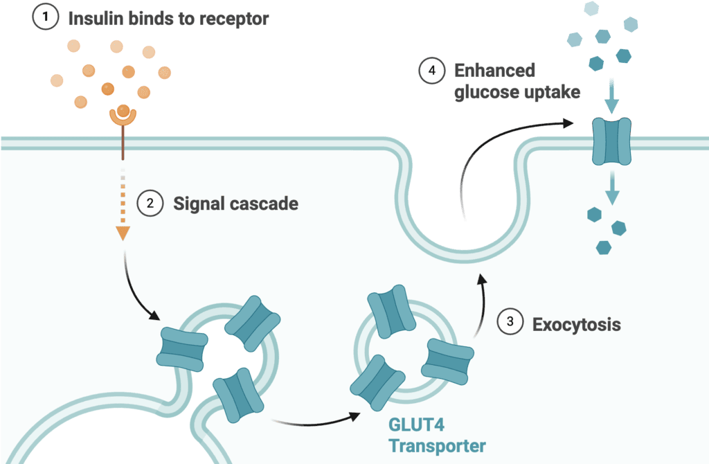
Figure 22. Insulin binding activates an intracellular signaling cascade that promotes translocation of GLUT4-containing vesicles to the plasma membrane. The resulting increase in surface GLUT4 transporters enhances glucose uptake by skeletal muscle and adipose tissue.
GLUT5 differs from the other major GLUT transporters because it primarily transports fructose rather than glucose. It is found mainly in the small intestine, where it facilitates fructose absorption from the intestinal lumen into enterocytes (intestinal/mucosal cells).
Figure 23. Approximate glucose affinity (Km) and transport capacity (Vmax) of the major GLUT transporters. GLUT3 exhibits the highest glucose affinity, whereas GLUT2 has the highest transport capacity, reflecting their specialized physiological roles.
Glucose Trapping Within Cells
Most tissues express an enzyme called hexokinase. This enzyme is well known because it also catalyzes the first step of glycolysis, which will be discussed in detail in a later post. Before glucose can participate in glycolysis or other metabolic pathways, however, it must first be retained within the cell. Because GLUT transporters mediate bidirectional facilitated diffusion, intracellular glucose could otherwise diffuse back out of the cell as concentrations equilibrate across the plasma membrane. Hexokinase prevents this through a process commonly known as glucose trapping.
Once glucose enters a cell, hexokinase rapidly phosphorylates it using the following reaction: Glucose + ATP → Glucose-6-phosphate + ADP
The addition of a phosphate group dramatically changes the properties of glucose. Unlike free glucose, glucose-6-phosphate cannot cross the plasma membrane and is not transported by GLUT proteins. As a result, phosphorylation traps glucose within the cell, keeping intracellular glucose concentrations low. This preserves the concentration gradient that favors continued glucose uptake while committing glucose to intracellular metabolism.
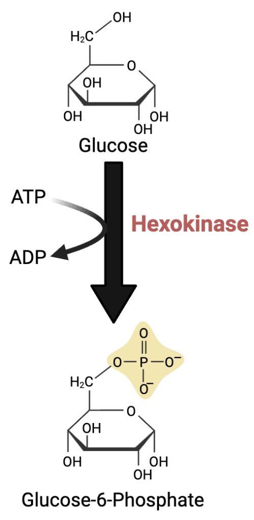
Figure 24. …
Although hexokinase is often discussed as a single enzyme, mammals actually express four different hexokinase isozymes (HK I-IV). While these isozymes differ in their tissue distribution and kinetic properties, they all catalyze the same reaction: the phosphorylation of glucose to glucose-6-phosphate. This reaction traps glucose inside the cell and commits it to cellular metabolism, allowing it to be used for pathways such as glycolysis, glycogen synthesis, and the pentose phosphate pathway. The differences between the individual hexokinase isozymes are important and will be explored in the next lesson.
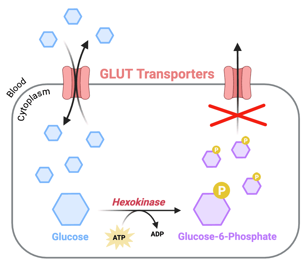
Figure 25. …
Conclusion
The journey of glucose from the diet to the interior of a cell involves a coordinated series of processes that include digestion, absorption, transport, distribution, and cellular uptake. Dietary carbohydrates are broken down into monosaccharides, absorbed by the small intestine, and delivered to the liver through the portal circulation. From there, glucose enters the bloodstream and is distributed to tissues throughout the body.
Specialized transport proteins allow glucose to enter different cell types according to their metabolic needs, while phosphorylation by hexokinase or glucokinase traps glucose within the cell and prepares it for metabolism. At this point, glucose is positioned to enter pathways such as glycolysis, where its stored chemical energy can be converted into ATP and other biologically useful forms of energy.
Quick Recap
- Glucose is a major metabolic fuel and an important precursor for the synthesis of many biomolecules.
- Dietary carbohydrates are digested into monosaccharides before they can be absorbed.
- The primary dietary carbohydrates include starch, glycogen, sucrose, lactose, maltose, and cellulose, each with distinct glycosidic linkages.
- Salivary and pancreatic α-amylases hydrolyze internal α(1→4) glycosidic bonds, while brush-border enzymes complete carbohydrate digestion into glucose, galactose, and fructose.
- Glucose and galactose are absorbed by SGLT1 through Na⁺-coupled secondary active transport, whereas fructose enters enterocytes through GLUT5 by facilitated diffusion.
- All absorbed monosaccharides leave enterocytes through GLUT2 and travel to the liver through the hepatic portal circulation.
- The liver serves as a metabolic buffer, helping regulate blood glucose by taking up or releasing glucose as needed.
- Insulin lowers blood glucose by promoting glucose uptake and storage, whereas glucagon raises blood glucose by stimulating hepatic glucose production and release.
- Different tissues express specialized glucose transporters (GLUT1-5) that match their metabolic demands.
- Once inside a cell, glucose is phosphorylated by hexokinase or glucokinase to form glucose-6-phosphate, trapping it inside the cell and preparing it for metabolism.