
Introduction to Metabolism
The Basis of Metabolic Biochem
Biochemistry is the study of the chemical processes that occur within living organisms. Fundamentally, biochemistry aims to describe how cells obtain and use energy, transform molecules, communicate with each other, and ultimately maintain life. Rather than focusing solely on tissues, organs, or entire organisms, biochemistry examines biology at the molecular level, where countless chemical reactions collectively determine cellular function. Understanding the connection between molecular structure and biological activity is one of the primary goals in biochemistry. In fact, one of the ideas I most vividly remember hearing throughout my undergraduate biochemistry courses was that structure determines function. The shape, bonding patterns, and chemical properties of a molecule largely dictate how it interacts with other molecules and, consequently, how cells behave, respond to signals, and carry out metabolism.
Metabolism refers to the complete collection of chemical reactions occurring within a cell or organism. These reactions are not isolated events but rather part of a highly coordinated and interconnected network that allows living systems to meet their energy and biosynthetic demands. From a thermodynamic perspective, living organisms continuously resist the natural tendency toward increasing disorder. Maintaining cellular organization therefore requires a constant input and utilization of energy. As a result, metabolism is essential for sustaining life. Despite the extraordinary diversity of organisms on Earth, many of the central metabolic pathways are highly conserved across species. This conservation reflects the fundamental importance of metabolism and makes it one of the major unifying principles of biology.
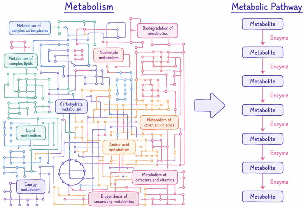
Metabolic reactions are organized into metabolic pathways, which consist of a series of interconnected enzymatic reactions that convert a starting molecule into a specific product. Each step within a pathway is catalyzed by a particular enzyme, making enzymes the primary functional units of metabolism. The molecules that participate in these reactions are known as metabolites. Because pathways proceed through a sequence of individual reactions, the product generated by one enzyme often serves as the substrate for the next. This stepwise organization allows cells to regulate metabolic activity with remarkable precision while maintaining both efficiency and adaptability.

Figure X. text
As you continue studying biochemistry, you will quickly discover that enzymes rarely work alone. Many enzymes require additional components known as cofactors to function properly. Cofactors are nonprotein molecules or ions that assist enzymes during catalysis and may be either organic or inorganic in nature. Organic cofactors are called coenzymes and are often derived from vitamins obtained through the diet. Coenzymes that bind temporarily to an enzyme, undergo chemical modification during the reaction, and are regenerated elsewhere are known as co-substrates. In contrast, coenzymes that remain tightly or permanently associated with an enzyme are called prosthetic groups. Inorganic cofactors commonly include metal ions such as magnesium, zinc, and iron, which may stabilize charges, facilitate substrate binding, or directly participate in catalytic mechanisms. Many biochemical reactions would proceed far too slowly, or would not occur efficiently at all, without the assistance of these essential molecules.

Figure X. text
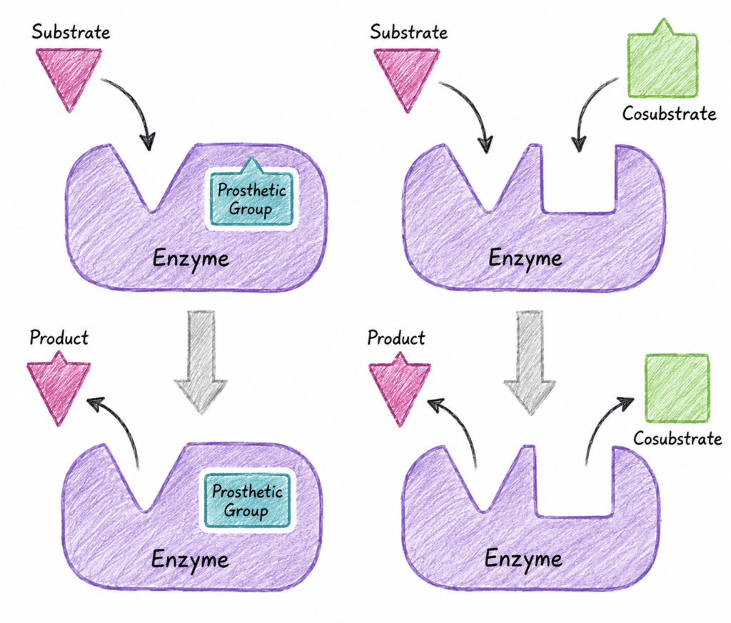
Figure X. text
Although metabolism encompasses thousands of individual reactions, most serve one or more of four major functions within the cell. First, metabolism captures and utilizes free energy to power cellular processes. Second, it degrades molecules when necessary to recover energy or recycle their components. Third, it converts nutrients into the building blocks required for cellular growth and maintenance. Finally, it assembles these building blocks into the complex macromolecules and organized structures that make up living cells. Together, these functions enable organisms to grow, maintain cellular integrity, reproduce, and respond to changes in their environment.
Figure X. text
Major Types of Metabolic Pathways
Metabolism is generally divided into three major categories: catabolism, anabolism, and amphibolic pathways. Catabolic pathways are responsible for the breakdown of larger molecules into smaller products. These reactions often involve the oxidation of nutrients and release free energy. Since they release energy, catabolic reactions are generally exergonic and have a negative Gibbs free energy change (ΔG < 0). Cells capture much of this energy in molecules such as ATP, NADH, and FADH₂, which can then be used to power other cellular processes. Major examples of catabolic pathways include glycolysis, β-oxidation, and glycogen degradation.
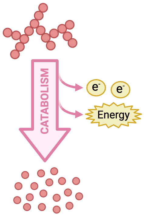
Figure X. text
Anabolic pathways are essentially the opposite. They involve the biosynthesis of larger, more complex molecules from smaller precursors. Since bond formation requires an energy input, anabolic reactions are generally endergonic and have a positive Gibbs free energy change (ΔG > 0). These pathways often rely on ATP and reducing power supplied primarily by NADPH. Anabolic pathways are responsible for synthesizing many of the molecules required for growth and cellular maintenance, including fatty acids, nucleotides, proteins, and carbohydrates.
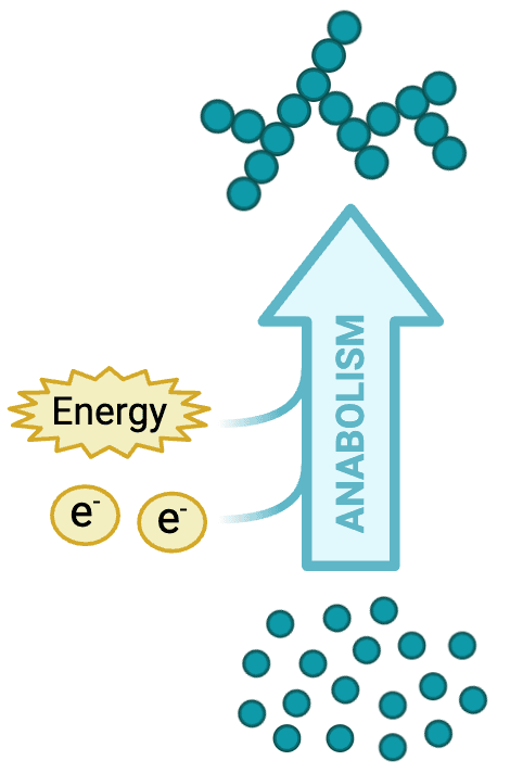
Figure X. text
Although catabolism and anabolism are often discussed separately, they are highly interconnected. Since my introductory chemistry and biology courses, one concept has been repeatedly emphasized: reactions cannot proceed without sufficient energy input. As a result, cells cannot build complex molecules unless enough energy has first been obtained elsewhere, meaning anabolism depends on the products of catabolism. Catabolic pathways provide both the energy and many of the metabolic intermediates required for anabolic processes. Consequently, metabolism functions as a highly integrated and coordinated network rather than a collection of independent reactions.
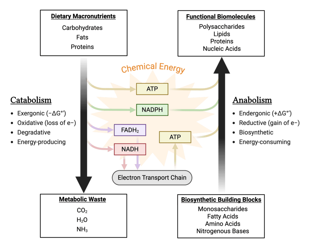
Figure X. text
Some pathways are considered amphibolic because they participate in both degradative and biosynthetic processes depending on cellular conditions. A classic example is the tricarboxylic acid (TCA) cycle, also known as the citric acid cycle or Krebs cycle. It functions catabolically by oxidizing acetyl-CoA for energy production, while also supplying intermediates used in biosynthetic pathways such as amino acid and heme synthesis. As a result, the TCA cycle plays an essential role at the center of metabolism and integrates numerous pathways throughout the cell.
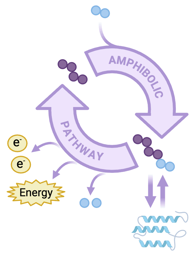
Figure X. text
This demonstrates one of the fundamental concepts in metabolic biochemistry: metabolic pathways do not work in isolation. Instead, they form a network of interconnected pathways that continuously exchange metabolites, electron/energy carriers, and biosynthetic intermediates. Recognizing these relationships provides a foundation for understanding metabolism as a whole and helps make sense of what can initially appear to be a complex web of chaos.
Thermodynamics & Free Energy
Laws of Thermodynamics
A fundamental concept behind metabolism is thermodynamics, which is the study of energy and its transformations. The First Law of Thermodynamics states that energy cannot be created or destroyed, only converted from one form to another. As a result, cells cannot generate energy from nothing. Instead, they capture energy from nutrients and convert it into forms that can be used to perform cellular work.
The Second Law of Thermodynamics states that the entropy, or disorder, of the universe tends to increase over time. At first glance, living organisms appear to contradict this principle because cells maintain a highly organized internal structure. However, cells do not violate the Second Law. The increase in order within a cell is accompanied by an even greater increase in disorder in the surrounding environment, largely through the release of heat during metabolic processes. Consequently, maintaining cellular organization requires a continual input and expenditure of energy.
Together, these thermodynamic laws provide the foundation for understanding energy transfer, reaction spontaneity, and metabolic organization within living systems.
Gibbs Free Energy
While the laws of thermodynamics describe the behavior of energy in general, biochemists often focus on a more practical quantity known as Gibbs free energy (ΔG). Gibbs free energy helps predict whether a reaction is thermodynamically favorable under a given set of conditions and is described by the following relationship: ΔG = ΔH − TΔS (see Equation 1).
ΔG = free energy change
ΔH = enthalpy (heat)
ΔS = entropy (disorder)
T = temperature (Kelvin)
Equation 1. Gibbs Free Energy.
Standard Free Energy Change
Because reaction conditions can vary considerably, biochemists often use a reference value known as the standard free energy change (ΔG°′). Under biochemical standard conditions, reactants and products are present at 1 M concentration, the pressure is 1 atm, the temperature is 25°C, and the pH is 7.0. Under these conditions, the relationship between free energy and equilibrium is described by: ΔG°′ = −RT ln (Keq)
This equation links the standard free energy change of a reaction to its equilibrium constant. Reactions with large equilibrium constants generally favor product formation and have negative ΔG°′ values, whereas reactions with small equilibrium constants tend to favor reactants.
ΔG = free energy change
ΔH = enthalpy (heat)
ΔS = entropy (disorder)
T = temperature (Kelvin)
Equation 2. Standard Free Energy Equation.
However, cells rarely exist under standard conditions. Metabolite concentrations are constantly changing as pathways consume reactants and generate products. As a result, the actual free energy change within a cell depends not only on ΔG°′ but also on the concentrations of reactants and products: ΔG = ΔG°′ + RT ln ([products]/[reactants])
This relationship helps explain why reactions that appear unfavorable based solely on their ΔG°′ values can still proceed within cells. If reactants are continuously supplied or products are rapidly removed, the actual ΔG can become negative, allowing the reaction to move forward.
ΔG = actual free energy change
ΔG°′ = standard free energy change
R = gas constant
T = temperature (Kelvin)
ln = natural logarithm
[P]/[R] = ratio of product to reactant concentration
Equation 3. Gibbs Free Energy Equation Under Cellular Conditions
I distinctly remember this being one of the first concepts in biochemistry that felt somewhat counterintuitive. Early on, it is easy to assume that a reaction with a positive ΔG°′ simply cannot occur. In reality, cells are dynamic systems, and metabolite concentrations play a major role in determining reaction direction. For this reason, the actual ΔG is often more informative than the standard free energy change when considering what is happening inside a cell.
This concept becomes especially important when studying metabolic pathways. The product of one reaction often becomes the reactant for the next, allowing cells to continuously influence reaction direction and control the flow of metabolites through a pathway.
ATP & Biological Energy
ATP Structure
After discussing free energy and reaction spontaneity, the next logical question is how cells actually capture and transfer energy. The primary answer is adenosine triphosphate (ATP), often referred to as the energy currency of the cell.
ATP consists of three major components: the nitrogenous base adenine, the five-carbon sugar ribose, and a chain of three phosphate groups. These phosphate groups are designated α, β, and γ moving outward from the ribose. The α-phosphate is attached to ribose through a phosphoester bond, whereas the β and γ phosphates are connected by phosphoanhydride bonds. These phosphoanhydride bonds have particularly large negative free energies of hydrolysis, making ATP an effective molecule for transferring energy between cellular processes.
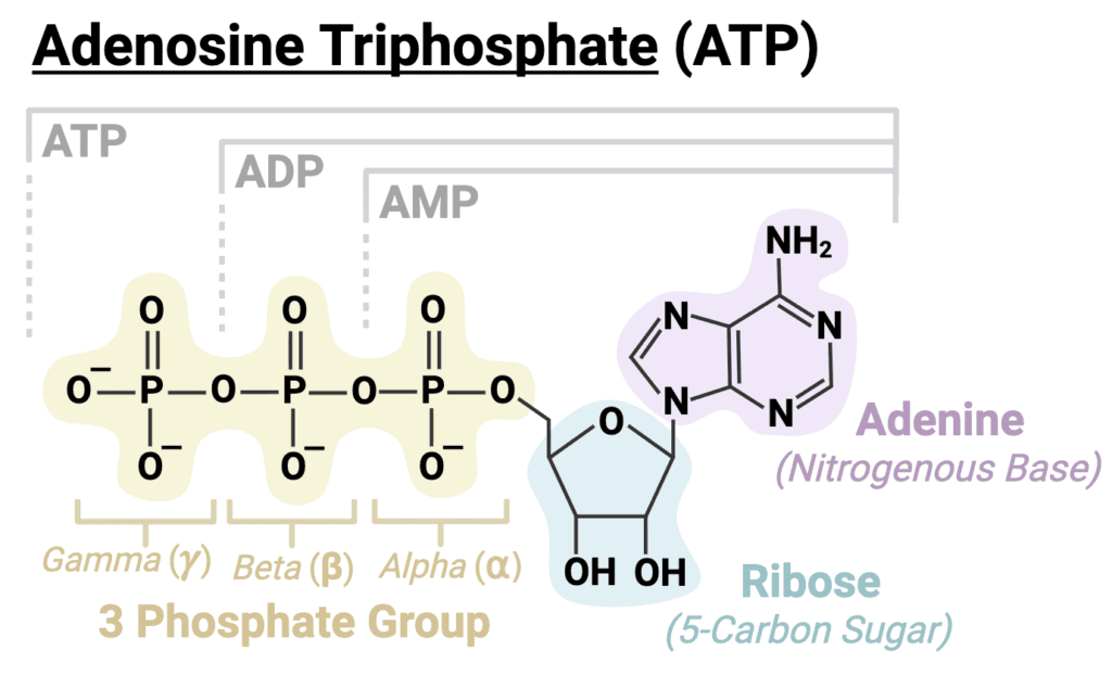
Figure X. text
ATP Hydrolysis & Energy Transfer
ATP is highly versatile because different phosphate bonds can be broken depending on the metabolic needs of the reaction. Different nucleophilic attacks on ATP produce different products and release different amounts of usable energy.
The most common form of ATP hydrolysis involves nucleophilic attack on the γ-phosphate, breaking the phosphoanhydride bond between the β and γ phosphates. This reaction produces ADP and inorganic phosphate (Pi) and is used extensively during phosphorylation reactions, enzyme regulation, active transport, and metabolic energy coupling.
ATP → ADP + Pi
A second mode of cleavage occurs when the bond between the α and β phosphates is broken. In this case, ATP is converted to AMP and pyrophosphate (PPi). Because pyrophosphate can subsequently be hydrolyzed to two inorganic phosphates, these reactions are often strongly favorable and are commonly used to drive biosynthetic processes.
ATP → AMP + PPi
A third mechanism involves transfer of an adenylyl group (AMP) to a substrate, producing an adenylated intermediate and releasing pyrophosphate. These activated intermediates play important roles in reactions such as aminoacyl-tRNA synthesis during protein translation and fatty acyl-CoA formation during lipid metabolism.
Although these reactions differ mechanistically, they all serve the same general purpose: coupling ATP hydrolysis to cellular processes that would otherwise be energetically unfavorable.
Why ATP Hydrolysis Is Favorable
A common misconception is that ATP releases energy simply because a phosphate bond is broken. In reality, breaking any chemical bond requires an input of energy. ATP hydrolysis is favorable because the products formed after hydrolysis are more stable than ATP itself.
Several factors contribute to this stability. First, ATP contains multiple negatively charged phosphate groups positioned close together, creating significant electrostatic repulsion. Hydrolysis relieves some of this repulsion. Second, the products of hydrolysis possess greater resonance stabilization than ATP. Finally, ADP and inorganic phosphate interact more favorably with surrounding water molecules, further lowering their free energy.
Taken together, these factors make the hydrolysis products more stable than ATP, resulting in a negative free energy change.
Figure X. text
Magnesium and ATP
Most ATP-dependent reactions require magnesium ions (Mg²⁺). Because ATP contains several negatively charged phosphate groups, free ATP is relatively unstable. Within cells, ATP is therefore usually present as a Mg-ATP complex rather than as free ATP. The positively charged magnesium ion helps shield negative charges on the phosphate groups, stabilizes the transition state, and assists with proper positioning of ATP within enzyme active sites.
Figure X. text
The ATP Cycle
Cells continuously cycle ATP between its high-energy and lower-energy forms. Energy released during catabolic pathways such as glycolysis, the TCA cycle, and oxidative phosphorylation is used to synthesize ATP from ADP and inorganic phosphate. ATP hydrolysis then powers processes such as biosynthesis, active transport, muscle contraction, and signal transduction.
This continual cycling of ATP represents one of the central organizing principles of metabolism. Catabolic pathways generate ATP, while ATP hydrolysis provides the energy needed for cellular work. In this way, ATP directly links energy-producing pathways with energy-consuming processes throughout the cell.
Figure X. text
Other High-Energy Compounds
ATP is the most familiar energy-transfer molecule in biology, but it is not the only high-energy compound found in cells. More broadly, high-energy compounds are molecules whose hydrolysis releases large amounts of free energy that can be used to drive otherwise unfavorable reactions.
A compound is generally considered high-energy if its hydrolysis releases an amount of free energy comparable to or greater than that released by ATP. Rather than allowing this energy to dissipate as heat, cells temporarily capture and transfer it through specialized molecules.
Several classes of biologically important high-energy compounds exist. These include pyrophosphates such as ATP, acyl phosphates such as 1,3-bisphosphoglycerate, enol phosphates such as phosphoenolpyruvate (PEP), thioesters such as acetyl-CoA, and guanidino phosphates such as phosphocreatine. Although their structures differ, each stores energy in a relatively unstable chemical arrangement that becomes more stable following hydrolysis.
Figure X. text
Not all high-energy compounds possess the same transfer potential. Some molecules have even more negative free energies of hydrolysis than ATP and can therefore drive ATP synthesis directly. Two important examples are phosphoenolpyruvate (PEP) and 1,3-bisphosphoglycerate, both of which participate in substrate-level phosphorylation. Phosphocreatine serves a different role, acting as a rapidly accessible phosphate reservoir that helps maintain ATP levels during periods of high energy demand in muscle.
Figure X. text
Another important high-energy molecule is acetyl-CoA. Unlike ATP, its energy is stored within a thioester bond rather than a phosphate bond. Because sulfur participates in resonance stabilization less effectively than oxygen, thioesters are relatively reactive and release substantial free energy upon hydrolysis. Acetyl-CoA therefore functions both as a carrier of activated acetyl groups and as a central metabolic intermediate linking carbohydrate, lipid, and amino acid metabolism.
Figure X. text
REDOX Reactions & Electron Carriers
Redox Reactions in Metabolism
ATP serves as the primary energy currency of the cell, but much of the energy used to generate ATP originates from the transfer of electrons. For this reason, oxidation-reduction reactions, commonly called redox reactions, are a central feature of metabolism. Most catabolic pathways harvest energy by removing electrons from nutrient molecules and transferring them to specialized electron carriers. Rather than releasing all of this energy at once as heat, cells capture it in a series of controlled steps.
Oxidation refers to the loss of electrons, whereas reduction refers to the gain of electrons. A commonly heard pneumonic to remember these terms is OIL RIG: Oxidation Is Loss, Reduction Is Gain.
Since electrons cannot exist freely in biological systems, oxidation and reduction always occur together. When one molecule loses electrons, another molecule must gain them. Throughout metabolism, carbon atoms from nutrients such as glucose and fatty acids are gradually oxidized, while electron carriers become reduced.
Figure X. text
One concept that helped me make sense of metabolism as a student was realizing that many pathways are essentially systems for moving electrons from one molecule to another. Once you begin tracking the flow of electrons rather than memorizing individual reactions, many metabolic pathways become much easier to understand.
The amount of energy stored within a molecule is closely related to its degree of reduction. Carbon-hydrogen bonds contain electrons with relatively high potential energy, so molecules that contain many carbon-hydrogen bonds are considered highly reduced. In contrast, carbon-oxygen bonds represent a more oxidized state because oxygen attracts electron density much more strongly than carbon. Carbon dioxide is the fully oxidized end product of carbon metabolism and contains very little usable energy. This is one reason why fatty acids, which contain many reduced carbon atoms, yield substantially more ATP upon complete oxidation than carbohydrates.
NAD⁺ and NADH
The primary electron carrier used in catabolic metabolism is nicotinamide adenine dinucleotide (NAD⁺). During oxidation reactions, NAD⁺ accepts two electrons and one proton to form NADH:
__
The transferred electrons are typically donated as a hydride ion (H⁻), which contains two electrons and one proton. NADH then carries these high-energy electrons to the electron transport chain, where their energy can ultimately be used to drive ATP synthesis.
Since NAD⁺ primarily accepts electrons generated during nutrient oxidation, it is most commonly associated with catabolic pathways such as glycolysis, pyruvate oxidation, the TCA cycle, and β-oxidation.
Figure X. text
NADP⁺ and NADPH
NADPH is structurally similar to NADH but serves a different metabolic function. While NADH is primarily involved in ATP production, NADPH is used mainly in reductive biosynthesis and cellular antioxidant systems.
Many anabolic pathways require a source of electrons to build complex molecules. NADPH provides these electrons during reactions such as fatty acid synthesis and cholesterol synthesis. Cells therefore maintain a relatively high ratio of NADPH to NADP⁺, ensuring that reducing power is readily available whenever biosynthetic reactions occur.
A useful distinction is that NADH is generally associated with energy production, whereas NADPH is generally associated with biosynthesis.
Figure X. text
FAD and FADH₂
Another important electron carrier is flavin adenine dinucleotide (FAD). Unlike NAD⁺, which accepts electrons as a hydride ion, flavin cofactors can participate in either one-electron or two-electron transfer reactions. This flexibility allows FAD-containing enzymes to catalyze reactions that NAD⁺ cannot readily perform.
When FAD accepts electrons, it is reduced to FADH₂. Flavin cofactors are particularly important in pathways such as β-oxidation, the TCA cycle, and the electron transport chain. In contrast to NAD⁺, which typically functions as a freely diffusible co-substrate, FAD is often tightly bound to the enzyme that uses it.
Figure X. text
Reduction Potential & Electron Flow
The tendency of a molecule to gain electrons is described by its reduction potential, denoted E°′. Molecules with more positive reduction potentials have a stronger tendency to accept electrons and become reduced, whereas molecules with more negative reduction potentials are more likely to donate electrons.
Electrons therefore flow spontaneously from molecules with lower electron affinity to molecules with higher electron affinity. The free energy associated with this electron transfer is described by the relationship:
Figure X. text
In this equation, n represents the number of electrons transferred and F represents Faraday’s constant. A positive ΔE°′ produces a negative ΔG°′, indicating that electron transfer is thermodynamically favorable.
One of the most important examples in metabolism is the transfer of electrons from NADH to oxygen during oxidative phosphorylation:
NADH + H⁺ + ½ O₂ → NAD⁺ + H₂O
This reaction releases a large amount of free energy because oxygen has an exceptionally high affinity for electrons. However, cells do not transfer electrons directly from NADH to oxygen. Instead, electrons pass through a series of intermediate carriers within the electron transport chain. By releasing energy in many small steps rather than a single large step, cells can capture a significant portion of that energy and use it to generate the proton gradient that drives ATP synthesis.
Figure X. text
As we move deeper into metabolism, electron flow becomes one of the most useful concepts for understanding pathway function. Many pathways that initially appear unrelated are ultimately connected by a common purpose: extracting electrons from nutrients and transferring them to their final destination.
Metabolic Regulation
Introductory Concepts
Pathway Regulation
One of the most important ideas in metabolism is that metabolic pathways are not simply collections of chemical reactions. Cells must constantly regulate which pathways are active, how quickly they operate, and whether nutrients should be broken down or stored. Because a cell’s needs are always changing, metabolism must remain carefully coordinated to maintain homeostasis and respond to changing conditions.
Not every reaction within a metabolic pathway contributes equally to this regulation. Some reactions can readily proceed in either direction depending on the concentrations of reactants and products. Others are strongly energetically favorable and are essentially irreversible under physiological conditions. These irreversible reactions often serve as the major regulatory points of a pathway because controlling these steps largely controls the overall activity of the pathway.
As you work through metabolism, you’ll find that cells regulate these important steps in several different ways. The major types of metabolic regulation will be introduced in the section below.
Metabolic Flux
Another important concept in metabolism is metabolic flux, which refers to the overall rate at which molecules move through a metabolic pathway. Rather than simply turning pathways completely “on” or “off,” cells continuously adjust how quickly pathways operate to match their current needs. Some pathways may run very rapidly, others more slowly, and some may become nearly inactive until they are needed again. Since the demands of a cell are constantly changing, metabolic flux is also constantly changing. Regulating pathway flux allows cells to use energy efficiently while maintaining a stable internal environment.
Understanding Metabolic Logic
As you study metabolism, you’ll notice that pathways become much easier to understand when you focus on the movement of carbon atoms and electrons, rather than trying to memorize every reaction. Tracking carbon atoms helps explain how molecules are rearranged as they move through different pathways, while tracking electrons helps explain how cells capture, transfer, and use energy. Understanding metabolism therefore involves much more than memorizing pathways. It means understanding why reactions occur, how pathways are regulated, and how all of the reactions within the cell work together as one coordinated system.
Major Types of Metabolic Regulation
Cells use many different mechanisms to regulate metabolism. Some adjust the activity of existing enzymes within seconds, while others produce longer-lasting changes by altering the amount of enzyme that is made. Rather than relying on a single regulatory strategy, cells combine multiple mechanisms to regulate enzyme activity, enzyme abundance, and overall metabolic flux. Together, these mechanisms ensure that metabolic pathways operate at the right place, at the right time, and at the appropriate rate. The following are some of the major ways metabolism is regulated.
Allosteric Regulation
Allosteric regulation occurs when a regulatory molecule binds to a site on an enzyme other than the active site, called the allosteric site. This changes the enzyme’s shape, making it either more or less active. Because enzyme activity can change almost immediately, allosteric regulation is one of the fastest and most common forms of metabolic control.
Figure X. text
Feedback Inhibition
Feedback inhibition occurs when a downstream product inhibits an earlier enzyme within the same metabolic pathway. This prevents unnecessary product accumulation once enough has been made and helps maintain metabolic balance. Feedback inhibition is commonly achieved through allosteric regulation, although product inhibition, covalent regulation, and other mechanisms can also contribute depending on the pathway.
Figure X. text
Feedforward Activation
Feedforward activation is the opposite of feedback inhibition. In this mechanism, an early metabolite activates a later enzyme in the pathway, allowing pathway activity to increase before later reactions become overloaded with intermediates. Like feedback inhibition, feedforward activation is often mediated through allosteric regulation.
Figure X. text
Product Inhibition
Product inhibition occurs when the product of a reaction inhibits the enzyme that produced it. This prevents unnecessary accumulation of metabolic intermediates and helps maintain metabolic balance. Depending on the enzyme, product inhibition may occur through competitive inhibition at the active site or through allosteric regulation.
Figure X. text
Substrate Availability
Some metabolic pathways are regulated simply by the availability of their substrates. When substrate concentration increases, reaction rates often increase. When substrate becomes limited, pathway activity slows. Unlike allosteric regulation, this mechanism depends on the amount of substrate available rather than the binding of a separate regulatory molecule. Changes in substrate availability can also shift reaction rates according to the principles of mass action.
Figure X. text
Regulation by Energy Status
Cells continuously monitor their energy supply using molecules such as ATP, ADP, and AMP. Changes in the concentrations of these molecules signal whether energy is abundant or limited, allowing ATP-producing and ATP-consuming pathways to adjust accordingly. These molecules can regulate metabolism by binding allosterically to certain enzymes or by serving as reactants or products in enzyme-catalyzed reactions, allowing enzyme activity and metabolic flux to respond to the cell’s energy needs.
Figure X. text
Regulation by Energy Status
Many metabolic reactions depend on electron carriers such as NAD⁺, NADH, NADP⁺, and NADPH. Changes in the balance of these molecules influence whether oxidation and reduction reactions can proceed efficiently. These electron carriers can regulate metabolism by binding allosterically to certain enzymes or by serving as reactants or products in enzyme-catalyzed reactions, allowing enzyme activity and metabolic flux to respond to the cell’s current redox state.
Figure X. text
Covalent Regulation
Covalent regulation changes enzyme activity by adding or removing chemical groups, most commonly phosphate groups. These reversible modifications can either activate or inhibit an enzyme, allowing metabolism to respond rapidly to changing cellular conditions.
Figure X. text
Hormonal Regulation
Hormones coordinate metabolism throughout the entire body by allowing different tissues to communicate with one another. Many hormones regulate metabolism by activating signaling pathways that ultimately modify enzymes through covalent regulation, although they can also influence enzyme location within the cell and long-term changes in gene expression.
Figure X. text
Gene Expression Regulation
Cells can also regulate metabolism by changing how much of a particular enzyme is produced. Although this process is much slower than directly regulating enzyme activity, it allows metabolism to adapt to long-term changes in nutrient availability, hormones, and other physiological conditions.
Figure X. text
Compartmentalization
Cells separate many metabolic pathways into different regions of the cell, called cellular compartments. Restricting where reactions occur helps control substrate access, prevents incompatible pathways from interfering with one another, and allows different metabolic processes to be regulated independently.
Figure X. text
Tissue-Specific Regulation
Different tissues often regulate the same metabolic pathway in different ways because they perform different physiological functions. Differences in enzyme expression, hormone responsiveness, and metabolic demand allow the same pathway to meet the specific needs of each tissue. Different tissues may also express different transporters and regulatory proteins that further adapt metabolism to their physiological roles.
Figure X. text
Isozyme Regulation
Isozymes are different forms of the same enzyme that catalyze the same reaction but differ in their kinetic or regulatory properties. By expressing different isozymes in different tissues, cells can regulate the same metabolic pathway according to each tissue’s specific physiological role.
Figure X. text
Reciprocal Regulation
Many metabolic pathways perform opposing functions. Reciprocal regulation ensures that when one pathway becomes more active, the opposing pathway is simultaneously suppressed. This prevents futile cycles, in which opposing pathways run at the same time and consume energy without producing any useful net result. It also allows cells to efficiently direct metabolites toward the pathway that is most needed under a given set of conditions.
Figure X. text